Search conditions, treatments, research reviews and articles. Press Enter to see all results, Esc to close.
Search conditions, treatments, research reviews and articles. Press Enter to see all results, Esc to close.
Blog
October 31, 2026
A labeled eye diagram shows the eye from the side, following light from front to back, and from the front, as you see it in a mirror. Light passes through the cornea, pupil and lens, then crosses the clear vitreous gel to the retina. The retina turns light into signals, and the optic nerve carries them to the brain [1].
This article is general education. Each part below is explained with what it does and the conditions that affect it. A diagram cannot show which part is causing your symptoms; only an eye exam can do that.
Key takeaways
In this article
Some eye problems cannot wait for a routine visit. If you have any of these symptoms now, act first and read later.
Netra’s guide to urgent vision changes lists more warning signs.
The main parts of the eye are the cornea, iris, pupil, lens, retina and optic nerve. They sit inside a tough white wall called the sclera. Two clear fluids fill the eye: watery aqueous humor in front of the lens and gel-like vitreous behind it. The eyelids, lashes and tears protect the surface [1,6].
The eye wall has three layers [6]:
The side view, also called a cross-section, shows the eye as if it were cut in half from front to back. It follows the path light takes, from the clear cornea at the front to the retina and optic nerve at the back. Figure 1 labels 15 parts, and each one is explained below in the same order.
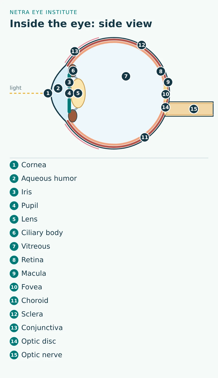
The cornea is the clear dome at the very front of the eye. It sits in front of the iris and pupil [12]. It does about two-thirds of the eye’s focusing, more than the lens [2].
The cornea has no blood vessels, which helps it stay clear. It is one of the most sensitive tissues in the body, which is why a scratch hurts so much [2]. Conditions of the cornea include infection or inflammation (keratitis), cone-shaped thinning (keratoconus), Fuchs dystrophy and corneal scratches.
Aqueous humor is a clear, watery fluid that fills the front of the eye, in front of the lens [6]. The space it fills, just behind the cornea, is called the anterior chamber [12]. The ciliary body makes this fluid all the time, and it drains out through tiny channels [13].
The balance between fluid made and fluid drained sets your eye pressure [14]. Many people with glaucoma have high eye pressure, and treatment that lowers it helps slow the disease [5]. Raised pressure without nerve damage is called ocular hypertension.
The iris is the colored part of the eye. It is a ring of muscle around the pupil. A muscle inside it narrows the pupil in bright light and when you focus up close. By changing the pupil’s size, the iris controls how much light reaches the retina [3]. The most common form of uveitis is inflammation of the iris [11].
The pupil is the round opening in the center of the iris. Light enters the eye through it [1]. The iris muscles make it smaller in bright light and larger in dim light [3].
The lens is a clear, flexible disc behind the iris. It hangs from fine fibers called zonules and changes shape to focus light on the retina [6,15].
With age the lens stiffens, so focusing up close gets harder. This usually starts in the early to mid-40s and is called presbyopia [15]. A cloudy area in the lens is a cataract. Cataracts are very common as people get older [16]. Netra’s guide to how lens clouding changes vision explains the stages.
The ciliary body is a ring of tissue just behind the iris with two jobs. Its muscle changes the shape of the lens so you can focus at different distances [15,17]. It also makes the aqueous humor [13].
The vitreous is the clear gel that fills the large space between the lens and the retina. It is about 98% water, held in a mesh of collagen and hyaluronic acid [18].
Early in life the vitreous is attached to the retina. With age it slowly separates. This is called posterior vitreous detachment. It often causes floaters and sometimes flashes. A sudden rise in floaters with flashes needs an eye exam right away, because the pull can tear the retina [18].
The retina is the thin layer of nerve tissue that lines the back of the eye [19]. Its photoreceptor cells turn light into electrical signals [1]. There are two kinds: about 92 million rods and about 4.6 million cones in an average retina [20].
Light passes through the whole thickness of the retina before it reaches the rods and cones. They sit at the back, against a pigment layer and the choroid [21]. Conditions of the retina include diabetic retinopathy, retinal vein occlusion and inherited diseases such as retinitis pigmentosa.
The macula is the small central part of the retina. It controls sharp, straight-ahead vision [22]. Damage here blurs the center of your vision, as in macular degeneration [22]. Swelling of the macula is macular edema.
The fovea is a small pit in the center of the macula. It is the spot where visual acuity, or sharpness, is highest [4]. Cones are packed most tightly here, and cone density drops steeply even 1 mm away [20]. A small break in this area is a macular hole.
The choroid is a layer full of blood vessels between the retina and the sclera [6]. It lies right under the retina’s light-sensing cells [21] and helps nourish the eye’s tissues [23]. Conditions that involve the choroid include central serous chorioretinopathy and choroiditis.
The sclera is the white of the eye. It is the tough, fibrous outer wall that protects the inside of the eye [6]. At the front it joins the clear cornea [12].
The conjunctiva is a thin, clear membrane. It lines the inside of the eyelids and covers the white of the eye. It makes mucus and tears that protect and lubricate the surface, helps keep germs out, and is rich in tiny blood vessels [7]. Loose folds of this layer can cause dry-eye symptoms, a condition called conjunctivochalasis.
The optic disc is the spot at the back of the eye where nerve fibers from the retina gather and leave the eye. Blood vessels also enter the retina here [21]. Eye doctors look at the disc during a dilated exam. Swelling of the disc is called papilledema.
The optic nerve carries visual signals from the retina to the brain, which turns them into the images you see [1,24]. It also carries the signal for the pupil’s light reflex [24]. Glaucoma damages this nerve [5]. Other conditions include optic neuritis and optic nerve atrophy; see Netra’s optic nerve guide.
The front view shows the eye as you see it in a mirror: mostly the surface of the eye and the parts that protect it. Figure 2 labels nine parts. The iris, pupil and sclera are explained in the side view above.
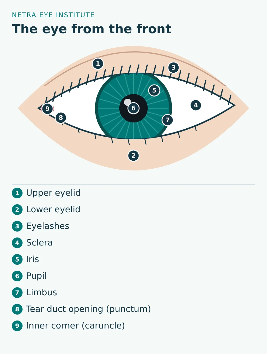
The eyelids spread tears across the eye, help drain tears and protect the surface. Their glands make oils that lubricate the eye [25]. These glands open along the lid edge and supply the oily top layer of the tears. Each blink renews the tear film and wipes away debris and germs [2].
An upper lid that droops is called ptosis. Lumps along the lid are often a chalazion or stye.
Eyelashes do real work. In wind-tunnel tests, lashes about one-third as long as the eye is wide halved both the dust reaching the eye and the drying of the tear film [26]. Lashes that turn inward and rub the eye cause a condition called trichiasis.
The limbus is the narrow border where the clear cornea meets the white sclera [12]. It holds stem cells that renew the surface of the cornea. When these cells are lost, eye surgeons can transplant small pieces of healthy limbus to restore the surface [27].
Near the inner corner, each eyelid has a tiny opening called a punctum. The plural is puncta. Tears drain into it, travel through small channels in the lids to the tear sac, and then flow into the nose [28]. If this drainage is blocked, tears spill over onto the cheek [29].
Over the whole front of the eye lies the tear film, a thin layer of oil, water and mucus [2]. When the eyes do not make enough tears, or tears dry up too fast, the result is dry eye [30]. Netra’s guide to the layers of the tear film explains how each layer works.
The caruncle is the small pink bump in the inner corner of the eye [31]. It is a patch of skin-like tissue with fine hairs, oil glands, sweat glands and a few extra tear glands [32]. It does not help you see. Growths here are uncommon, and 95% of 82 caruncle growths in one hospital series were benign. A new or changing bump should still be checked [32].
Light enters through the cornea, which does most of the focusing. The iris sets the size of the pupil to control how much light gets in. The lens fine-tunes the focus. Light then crosses the vitreous to the retina, where photoreceptors turn it into electrical signals. The optic nerve carries those signals to the brain, which builds the image [1,2].
If the eyeball is too long or too short, or the cornea is unevenly curved, light does not focus on the retina and vision blurs [33]. In nearsightedness, or myopia, light focuses in front of the retina [34]. Uneven corneal curve causes astigmatism [33]. Netra’s guide to how the eye and brain create vision picks up the story from the optic nerve onward.
Most eye conditions start in one main part of the eye. Dry eye affects the tear film, cataract the lens, glaucoma the optic nerve, macular degeneration the macula and diabetic retinopathy the blood vessels of the retina [5,8,16,22,30]. The table below matches common conditions to the part they affect.
| Part of the eye | Main job | Common conditions | Learn more |
|---|---|---|---|
| Tear film | Smooth, wet surface for clear vision | Dry eye | Dry eye |
| Cornea | Main focusing surface; protection | Keratitis, keratoconus, Fuchs dystrophy, scratches | Keratitis |
| Iris and middle layer (uvea) | Controls pupil size; blood supply | Uveitis | Uveitis |
| Aqueous humor and drainage | Fills the front of the eye; sets eye pressure | Glaucoma, ocular hypertension | Glaucoma |
| Lens and ciliary body | Fine focus, near focus | Cataract, presbyopia | Presbyopia |
| Vitreous | Clear gel that fills the eye | Floaters, posterior vitreous detachment | Floaters |
| Retina | Turns light into signals | Diabetic retinopathy, vein occlusion, retinitis pigmentosa, detachment | Diabetic retinopathy |
| Macula and fovea | Sharp central vision | Macular degeneration, macular edema, macular hole | Macular degeneration |
| Choroid | Blood vessels under the retina | Central serous chorioretinopathy, choroiditis | Choroiditis |
| Optic nerve | Carries signals to the brain | Glaucoma, optic neuritis, optic nerve atrophy | Optic nerve damage |
| Eyelids and lashes | Protect the surface; spread tears | Ptosis, chalazion, trichiasis | Ptosis |
A diagram shows where each part sits, not what is wrong with your eye. Blurry vision can come from the tear film, cornea, lens, shape of the eye, retina or optic nerve. These causes can feel much the same. Telling them apart takes an eye exam, often with dilating drops [5,33].
Diagrams are also simplified and not to scale. The cornea and retina are each only about half a millimeter thick [2,21], and parts drawn as one line hold several layers of cells. Netra’s guide to how often to have an eye exam explains what a full exam checks.
Netra Eye Institute, in South Plainfield, New Jersey, offers Netra Restoration Therapy (NRT). NRT is adjunctive care: it is used alongside the diagnosis, monitoring and treatment your eye doctor provides, never instead of them [35]. NRT does not change the structure of any part of the eye shown here. It has not been tested in controlled trials for the conditions named on this page.
If you are considering NRT, keep all standard treatment and follow-up in place, never delay care for an urgent symptom, and bring your recent eye exam reports. See NRT effectiveness by diagnosis for how Netra reviews outcomes.
There is no fixed number, because each part can be split into smaller ones. The diagrams on this page label 15 parts in the side view and nine in the front view. The core parts are the cornea, iris, pupil, lens, retina, macula, optic nerve, sclera, vitreous and aqueous humor.
The white part of the eye is called the sclera. It is the tough outer wall that protects the inside of the eye. A thin, clear layer called the conjunctiva covers it.
Light passes through the tear film, cornea, aqueous humor, pupil, lens and vitreous before it reaches the retina. It then passes through the retina’s inner layers to reach the rods and cones at the back. The optic nerve carries the resulting signals to the brain.
The cornea does most of the focusing, about two-thirds of the eye’s total focusing power. The lens behind the iris does the rest and fine-tunes focus for near and far. That is why changes in the shape of the cornea, such as astigmatism or keratoconus, can blur vision so much.
The small pink bump in the inner corner of the eye is the caruncle. It is a patch of skin-like tissue with fine hairs and tiny oil, sweat and tear glands. It does not help you see, and a new or changing bump there should be checked by an eye doctor.
Glaucoma damages the optic nerve, the cable that carries signals from the retina to the brain. Many people with glaucoma have high eye pressure, which builds when the fluid in the front of the eye does not drain well. Lowering eye pressure helps slow the damage.
A cataract affects the lens, the clear disc behind the iris that fine-tunes focus. A cataract is a cloudy area in the lens that makes vision blurry or dim. Cataracts are very common as people get older.
Sources checked October 31, 2026.
This article provides general education and does not replace an individual eye examination, diagnosis or treatment plan. If you notice a sudden burst of new floaters, flashes of light, a curtain over your vision or a painful red eye, get emergency eye care right away.