Search conditions, treatments, research reviews and articles. Press Enter to see all results, Esc to close.
Search conditions, treatments, research reviews and articles. Press Enter to see all results, Esc to close.
Blog
October 27, 2026
The optic nerve is the cable of nerve fibers that carries what your eye sees to your brain. Each one holds about 1.2 million fibers, which grow out of cells in the retina called retinal ganglion cells. Fibers that die do not grow back, so vision lost to glaucoma and most other optic nerve damage stays lost. Some causes, such as optic neuritis, often get better on their own, and early treatment protects the fibers that are left.
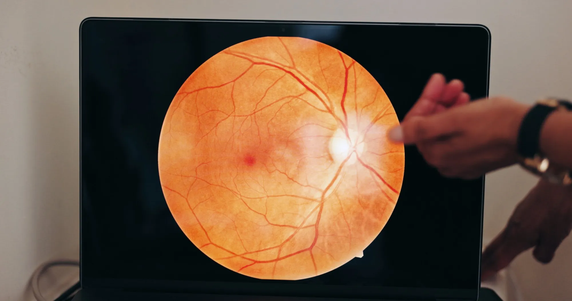
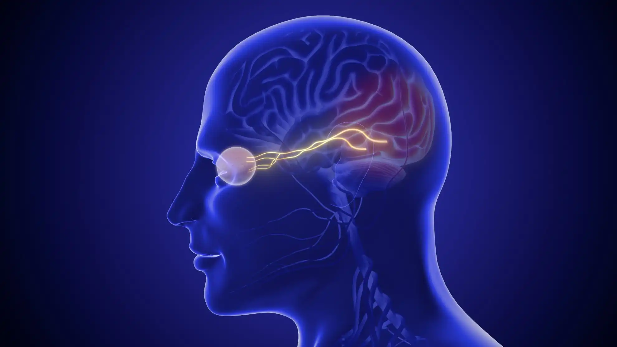
Light that enters the eye reaches the retina, which turns it into nerve signals. Those signals pass to retinal ganglion cells, whose long fibers gather at the optic disc, the round spot at the back of the eye, and leave the eye together as the optic nerve. In a study of 22 human optic nerves, the average count was 1,159,000 fibers, with a range of 816,000 to 1,502,000. The same study found that people lose about 5,400 fibers a year as they age.
On its way to the brain, the nerve runs about 1 mm inside the eye, 25 mm through the eye socket, 9 mm through a narrow bony canal and 16 mm inside the skull. The two optic nerves meet behind the eyes at the optic chiasm. There, the fibers from the inner half of each retina cross to the other side. That is why a pituitary tumor pressing on the chiasm can take away the outer (side) vision of both eyes.
The optic nerve does more than carry images. Its signals also control how your pupils react to light, help your eyes focus and set your body clock. Because it is part of the brain's wiring, it behaves like the brain and spinal cord: once its fibers are lost, they do not grow back. For a plain-English tour of the whole pathway, see how the eye and brain create vision.
Doctors call any optic nerve damage an optic neuropathy. The cause decides how fast it happens, what you notice and whether vision can come back.
| Cause | What happens and what you may notice | Can vision recover? |
|---|---|---|
| Glaucoma | Slow loss of nerve fibers, often with high eye pressure. In normal-tension glaucoma, it happens with normal pressure. The common open-angle type has no symptoms at first, and side vision fades slowly. Angle-closure glaucoma can start suddenly with intense eye pain, a red eye, blurred vision and nausea, and is a medical emergency. | No. Treatment can often stop or slow further loss |
| Optic neuritis | Inflammation of the nerve, sometimes the first sign of multiple sclerosis (MS). Pain when moving the eye, blurred or dim vision in one eye over hours or days, faded colors. | Usually. Most people recover over weeks to months, but some loss can be permanent |
| NAION (non-arteritic anterior ischemic optic neuropathy) | A drop in blood flow to the front of the nerve. Sudden, painless vision loss in one eye. | Sometimes partly. No treatment is proven; in the main trial, about 4 in 10 people who had no surgery could read 3 or more extra lines on the eye chart after six months |
| Giant cell arteritis | Inflamed arteries cut off the nerve's blood supply, mostly in people over 50. Vision loss, often with a new headache, scalp tenderness or jaw pain when chewing. | Lost vision rarely returns. Fast steroid treatment helps prevent further loss |
| Papilledema | Swelling of both optic discs from raised pressure inside the skull, for example from a tumor, a blood clot or idiopathic intracranial hypertension (raised pressure with no clear cause). Headaches, brief blackouts of vision lasting seconds, double vision, or no symptoms. | The outlook is good if it is found early; untreated, it can cause blindness |
| Compression | A tumor, such as a pituitary tumor or a meningioma (a usually noncancerous tumor of the brain's lining), a bulging blood vessel (aneurysm) or thyroid eye disease presses on the nerve. Slow vision loss, often with headaches. | Sometimes, if the pressure is removed before the nerve wastes away |
| Medicines and toxins | Ethambutol, a tuberculosis medicine, affects about 1 in 100 people who take it at standard doses. Methanol poisoning can also damage the nerve. | Ethambutol damage is largely preventable with regular checks. After methanol poisoning, 30% to 40% of survivors keep lasting vision problems |
| Poor nutrition | A lack of certain vitamins and nutrients, for example after weight-loss surgery. | Can be excellent if the missing nutrients are replaced in time |
| Inherited disease | Leber hereditary optic neuropathy (LHON) comes from a change in mitochondrial DNA, the genes in the cell's energy-making parts, passed down from the mother. It usually starts in young adults, men are four to five times more likely to lose vision, and painless central vision loss in one eye is followed by the other weeks to months later. | Treatment options are limited; in the EU, idebenone is approved for vision loss from LHON. Carriers who smoke are much more likely to lose vision |
| Injury | Traumatic optic neuropathy after car or bicycle accidents, falls, sports injuries or assaults. Vision loss, blur or faded colors. | Some vision often returns on its own. Steroids and surgery have not been shown to help and carry risks, so treatment is decided case by case |
Less common causes include optic nerve drusen (deposits in the optic disc), optic nerve tumors such as gliomas (tumors of the nerve's support cells) and meningiomas, neuromyelitis optica spectrum disorder (an immune condition that inflames the optic nerves and spinal cord) and conditions present from birth, such as optic nerve coloboma (a gap in the nerve's structure). Damage usually changes how the optic disc looks. In glaucoma, its center hollows out, which doctors call cupping. In most other causes, the disc turns pale. This is called optic nerve atrophy, and once it is established, vision rarely comes back.
Semaglutide has been linked to NAION. In June 2025, the European Medicines Agency concluded that NAION is a very rare side effect of semaglutide (Ozempic, Rybelsus and Wegovy), affecting up to 1 in 10,000 users. If you take semaglutide and notice sudden vision loss, get emergency care and tell the doctor who prescribes it.
The most common type of glaucoma, open-angle glaucoma, usually causes no symptoms early on, which is why regular eye exams matter. Other optic nerve problems can cause:
Go to an emergency room now if: you suddenly lose vision in one or both eyes, even if it comes back; you have intense eye pain with a red eye, blurred vision or nausea, which can be angle-closure glaucoma; you have brief blackouts of vision with headaches; you notice a sudden shower of new floaters, flashes of light, or a shadow or curtain over part of your vision, which can be a retinal detachment; or your vision changes after an injury to your eye or head. Call 911 if vision loss comes with weakness, numbness, a drooping face or trouble speaking. Sudden vision loss can be an eye stroke, which is treated as a medical emergency.
Get medical help the same day if: your vision gets worse and it hurts to move your eye, or you are over 50 and have vision changes with a new headache, scalp tenderness or jaw pain when chewing. See when an eye problem is an emergency.
An eye doctor can see many optic nerve problems during an eye exam. Depending on the cause your doctor suspects, the checks can include:
Our guide to the glaucoma test explains these checks in more detail.
Not once its fibers have died. A 2015 NEI report on regenerating the optic nerve notes that humans and other mammals normally show no regrowth of these fibers, which explains why the vision loss is permanent. Researchers have tested many possible treatments in animals, but none has yet been shown to work in people.
What helps depends on the cause:
Researchers are also testing ways to protect stressed nerve cells. Nicotinamide, a form of vitamin B3, has shown early signals in small glaucoma trials, and larger trials are under way. One trial participant had liver injury likely caused by high-dose nicotinamide, so it should be taken only within a supervised trial. Gene therapy for LHON is being tested in clinical trials. Our guide to optic nerve damage covers recovery by cause and the newest treatments.
No food or supplement is proven to protect the optic nerve in people. For the evidence on eye supplements, see eye vitamins.
Netra Restoration Therapy (NRT) is an integrative program at Netra Eye Institute. For people with optic nerve conditions, it can include acupuncture, traditional Chinese medicine, Ayurvedic therapies and nutrition guidance. These are offered as supportive care, after the cause has been diagnosed and alongside your eye doctor's treatment. NRT does not treat glaucoma, lower eye pressure, regrow the optic nerve or restore lost vision, and none of these therapies has been proven to protect the optic nerve. It never replaces urgent care for sudden vision loss. Tell your eye doctor about any herbs or supplements you take, because some can interact with medicines and some botanical products can harm the liver. Read what the research shows on acupuncture for eye disease and Ayurvedic care for optic nerve damage, or learn about Netra Restoration Therapy.
The optic nerve is the nerve that carries visual signals from the retina at the back of your eye to your brain. It is a bundle of about 1.2 million fibers, roughly 50 mm (2 inches) long. Its signals also control your pupils and help set your body clock.
Common symptoms are blurred or dim vision, faded colors, blind spots, loss of side vision, pain when moving the eye and sudden vision loss. Open-angle glaucoma, the most common type, usually causes no symptoms at first, so regular eye exams are the way to catch it early.
Fibers that have died cannot be brought back yet, so vision lost to glaucoma or optic nerve atrophy is usually permanent. Treatment can often stop further damage, and some causes get better: most people recover good vision after optic neuritis, and removing pressure from a compressed nerve before it wastes away can improve sight.
Glaucoma and optic neuritis are two of the best known. Glaucoma slowly destroys nerve fibers, often without symptoms, while optic neuritis is an inflammation that usually causes painful vision loss in one eye and often improves. Others include NAION, giant cell arteritis, papilledema and LHON.
An optometrist can. In the UK, 'the opticians' is where an optometrist does your eye test, and the NHS says glaucoma is usually found at a routine eye test, often before symptoms start. In the US, an optician fits glasses but does not check eye health; an optometrist or ophthalmologist checks your optic nerve. If damage is found, you may be referred to an ophthalmologist.
A pale optic disc usually means optic nerve atrophy: the nerve has lost many fibers. It is not a disease in itself but the result of an earlier cause, such as poor blood flow, inflammation, compression, a toxin or an inherited condition. Glaucoma more often hollows the disc out than turns it pale. Your doctor will look for the cause, because some need urgent treatment.
No food, supplement or exercise has been proven to strengthen the optic nerve. The steps that protect it are regular eye exams, taking glaucoma treatment as prescribed, managing diabetes and blood pressure, and not smoking if you carry an LHON gene change. Nicotinamide is being studied for glaucoma, but high doses have caused liver injury, so take it only within a supervised trial.
Medical Disclaimer: This article provides general education. It is not medical advice, a diagnosis or a personal treatment plan, and it does not establish a doctor-patient relationship. Do not start, stop or change any prescribed medicine, supplement or treatment without the clinicians responsible for your care. Sudden vision loss, or intense eye pain with a red eye, needs emergency care now. Vision that gets worse with pain on eye movement, or vision changes with a new headache, scalp tenderness or jaw pain, needs same-day medical care.