Blog
August 9, 2026
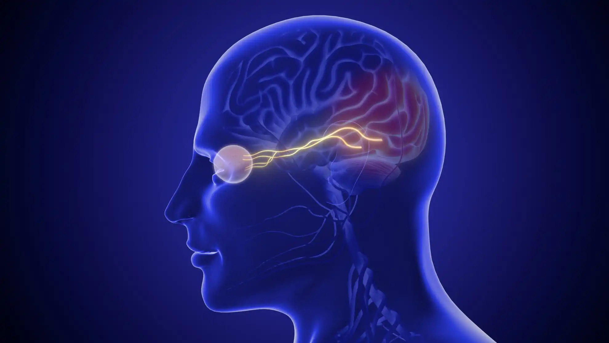
Your eyes collect light; your brain builds the picture. The retina converts photons into signals, the optic nerves carry them to the visual cortex at the back of the head, and the brain fills gaps, steadies movement and supplies color and meaning. This page traces that path and where it can break down.
Seeing feels instantaneous. Light enters the eyes and a meaningful world appears: edges, color, distance, motion, faces and words. Behind that apparent simplicity is a chain of optics, cellular signaling and neural interpretation. A clear image must first be formed on the retina, but the retina does more than record a picture. It begins processing contrast, color, movement and timing before signals ever reach the brain.
Understanding this system makes many eye-care terms easier to interpret. Nearsightedness concerns where light focuses. Cataract reduces optical clarity. Macular disease affects detailed central retinal function. Glaucoma and other optic neuropathies damage the nerve pathway. Stroke or traumatic brain injury can disrupt visual fields, attention or interpretation even when the eyes themselves appear healthy.
This guide follows vision from the tear film to the visual brain, explains what common tests measure and clarifies where medical treatment, optical correction and rehabilitation have different roles.
Before light can be focused, it must pass through a stable tear film. Tears are not simply water. They contain a complex mixture of lipids, aqueous fluid, mucins, proteins and immune components. The outer lipid layer slows evaporation, aqueous tears supply moisture and dissolved substances, and mucins help tears spread across the corneal surface.
Each complete blink redistributes the film and clears small debris. When blinking becomes less frequent or incomplete—often during concentrated screen use—the optical surface can break up between blinks. The result may be fluctuating blur, burning or glare that briefly improves after blinking. This is why dry-eye symptoms can affect visual quality even when the internal structures are healthy.
Eyelids, lashes, the bony orbit and protective reflexes reduce injury. The conjunctiva lines the inner eyelids and covers the visible sclera, while the sclera forms the strong white outer wall of the globe. These structures support and protect vision but are not passive; inflammation, allergy, infection and eyelid-position problems can disrupt the ocular surface.
The cornea is the transparent dome at the front of the eye. Light changes direction when it passes between materials with different refractive properties. Because the air-to-cornea boundary creates a strong change, the cornea supplies most of the eye’s fixed focusing power.
A smooth, regularly curved cornea helps rays converge. If curvature is too steep, too flat or uneven, the retinal image blurs. Astigmatism describes unequal focusing in different meridians. Keratoconus and corneal ectasia create more irregular distortion that ordinary spherical glasses may not fully correct. Scarring, swelling and surface disease can scatter light and reduce contrast.
The cornea has no blood vessels in its central optical zone. It obtains oxygen and nutrients through tears, the aqueous humor and surrounding tissues. Dense sensory nerves make it exquisitely sensitive. That sensitivity helps trigger blinking and tearing, but it also explains why a small abrasion can be extremely painful.
The iris is the colored ring seen through the cornea. Muscles within it change the diameter of the pupil, the central opening. In bright light the pupil usually constricts; in dim light it enlarges. Pupil size also changes with near focus, emotion, medications and neurologic activity.
The pupil helps balance sensitivity and optical quality. A smaller pupil can increase depth of focus and reduce some peripheral aberrations, which is why a pinhole may temporarily sharpen certain refractive blurs. A very large pupil admits more light in darkness but also exposes more of the optical system’s imperfections.
Pupil responses provide information about the retina, optic nerve, brainstem and autonomic pathways. A new unequal pupil, especially with drooping eyelid, double vision, pain or neurologic symptoms, needs prompt assessment. The pupil itself does not “see”; it controls how much light enters.
Behind the iris sits the crystalline lens. It is suspended by zonular fibers connected to the ciliary body. For near vision, the ciliary muscle changes tension so the lens becomes more curved and increases focusing power. This process is accommodation.
Young lenses are flexible. With age they become less able to change shape, leading to presbyopia—the progressive need for additional near focusing power. Presbyopia is a normal optical aging change, not a disease and not evidence that near work has weakened the eye.
The lens must also remain transparent. Cataract occurs when lens proteins and structure become cloudy enough to interfere with vision. Symptoms may include glare, haze, reduced contrast, altered color and prescription change. Glasses can address refractive error around an early cataract, but they cannot make an opaque lens transparent. When cataract meaningfully limits function, surgery replaces it with an artificial intraocular lens.
Accommodation works together with convergence, the inward rotation of both eyes for near targets, and with pupil constriction. These linked responses are coordinated by the nervous system. A person can have clear monocular optics yet experience fatigue or double vision when eye teaming is inefficient.
The large space behind the lens contains vitreous, a transparent gel composed mostly of water with a collagen and hyaluronan framework. It helps maintain shape and transmits light to the retina. With age, the gel becomes more liquefied and can pull away from the retina in a posterior vitreous detachment.
Condensations within the vitreous cast moving shadows perceived as floaters. A gradual, stable floater may be benign. A sudden shower of new floaters, flashes or a curtain-like shadow can indicate a retinal tear or detachment and requires immediate dilated evaluation. The symptom arises in the visual field, but the important event is physical traction at the retina.
The retina is layered neural tissue lining the back of the eye. Light passes through inner retinal layers before reaching photoreceptor outer segments. Photopigments change configuration when they absorb photons, beginning phototransduction—a biochemical process that changes the cell’s electrical activity.
Signals move through a network of bipolar, horizontal, amacrine and ganglion cells. Horizontal and amacrine circuits help compare information across space and time. Ganglion-cell axons gather at the optic disc and form the optic nerve. The retina therefore performs substantial preprocessing rather than sending an unedited camera image.
The retinal pigment epithelium beneath the photoreceptors absorbs stray light, participates in the visual cycle, transports nutrients and waste and renews photoreceptor outer segments. The choroid supplies a rich vascular bed behind it. Disorders involving photoreceptors, pigment epithelium, retinal vessels or supporting tissue can produce different patterns of visual loss.
Cones function best in brighter conditions and support color discrimination and high spatial detail. Human color vision depends on cone classes with different spectral sensitivities. They are densely packed in the fovea, the center of the macula.
Rods are more sensitive to low light and are numerous outside the foveal center. They do not provide normal color vision but support dim-light detection and peripheral awareness. Dark adaptation takes time because retinal sensitivity and photopigment states change after moving from brightness to darkness.
Night blindness can result from retinal disease, vitamin A deficiency, cataract and other conditions; it should not be assumed to be normal aging. Difficulty moving between lighting levels can also arise from reduced retinal function or optical scatter.
The macula is a specialized central retinal region. At its center, the fovea contains a high density of cones and neural wiring suited to fine resolution. Reading small print, recognizing faces and inspecting detail depend heavily on this area.
Macular disease often causes distortion, central blur or a missing central spot while peripheral vision remains. That pattern differs from glaucoma or retinitis pigmentosa, which may initially affect peripheral fields. “Retinal disease” is therefore not one uniform experience.
Peripheral vision is less detailed but highly important for detecting motion, monitoring the environment, balance and navigation. A person with excellent central acuity can still have disabling peripheral-field loss. Standard reading charts do not measure this function well; perimetry or confrontation field testing may be needed.
Photoreceptors do not send a separate full-resolution message for every point of light. Retinal circuits combine and compare signals. Receptive fields emphasize differences between neighboring regions, helping encode edges and local contrast. Some ganglion-cell pathways respond strongly to brightness differences, others to color relationships, timing or direction of motion.
This organization makes communication efficient. The optic nerve has far fewer fibers than the number of photoreceptors, so retinal networks compress information into patterns the brain can use. It also means retinal disease can alter quality in ways a simple blur analogy misses. A person may retain the ability to identify a high-contrast letter yet lose sensitivity to low contrast, motion or dim light.
Visual adaptation continually recalibrates sensitivity. In bright conditions, the system reduces gain so it is not overwhelmed. In darkness, rods and neural circuits become more sensitive over minutes. This is why walking from sunlight into a dark theater initially feels difficult. Cataract, retinal disease and aging can slow or limit adaptation.
Color perception is relational as well. Cone signals are compared through opponent channels rather than read as three independent colored pixels. Optic-nerve disease can reduce color saturation, sometimes more in one eye. Lens yellowing and filters alter the spectrum reaching the retina. Color complaints therefore require context: congenital difference, optical filtering, retinal change, optic neuropathy or medication can produce distinct patterns.
The inner retina is supplied by retinal vessels, while the photoreceptor-rich outer retina depends heavily on the choroid. The foveal center is relatively free of retinal capillaries to preserve optical clarity. Oxygen and nutrients must be delivered precisely because photoreceptors consume substantial energy.
Tight cellular junctions form inner and outer blood-retinal barriers that regulate movement of fluid and molecules. Diabetes, inflammation and vascular occlusion can disrupt these barriers, producing leakage and macular edema. Anti-VEGF medicines, corticosteroids, laser or surgery may be used depending on cause; glasses cannot remove retinal fluid.
Retinal vessels are directly visible during examination, giving clinicians a unique window into microvascular health. Findings can reflect diabetes, hypertension, inflammation or embolic disease, but an eye image is not a stand-alone diagnosis of whole-body health. Abnormalities must be coordinated with medical evaluation.
More than a million retinal ganglion-cell axons leave through the optic disc to form each optic nerve. The point where the nerve exits contains no photoreceptors, creating a normal blind spot that the brain usually makes unnoticed through binocular overlap and perceptual completion.
Fibers partially cross at the optic chiasm. Information from the right side of visual space is routed toward the left cerebral hemisphere and information from the left visual space toward the right. This organization explains why a brain lesion behind the chiasm often removes the same side of the visual field from both eyes.
The optic nerve can be injured by glaucoma, inflammation, ischemia, compression, toxic or nutritional factors, inherited disease and trauma. Because it is central nervous system tissue, substantial axonal loss is not restored by glasses. Treatment targets the cause and protects remaining function; rehabilitation may help a person use that function more effectively.
Most retinal output passes through the lateral geniculate nucleus of the thalamus before reaching primary visual cortex in the occipital lobe. From there, distributed networks analyze form, color, motion, depth, faces, objects, spatial relationships and visually guided action.
Vision is therefore an active inference. The brain combines input from two moving eyes with memory, attention and context. It stabilizes the scene during eye and head movements, fills normal gaps and selects information relevant to a task. Optical illusions expose some of these rules, but the same processes ordinarily make perception fast and efficient.
Broadly described dorsal networks contribute to spatial relationships and action—often summarized as “where/how”—while ventral networks contribute to object and identity recognition—often summarized as “what.” Real processing is interconnected rather than two simple channels.
A stroke, concussion or neurodegenerative disease can disrupt visual attention, field awareness, motion tolerance, reading or recognition. The eye examination may look normal because the difficulty lies farther along the pathway. Conversely, poor retinal input can increase the processing effort required from an intact brain.
Not every retinal signal reaches conscious awareness. Attention selects locations and features relevant to a goal. A person can look directly at a crowded shelf yet fail to find an item because search strategy, attention and target representation matter. Fatigue and cognitive load can reduce performance without changing the optical image.
After right-hemisphere stroke, some people develop left neglect: reduced awareness of the left side of space that is not explained fully by the eye’s visual field. Rehabilitation for neglect differs from treatment for a retinal defect, illustrating why localization matters.
Higher visual networks link shapes with stored knowledge. Damage can impair recognition of objects or faces despite adequate acuity. Reading similarly combines accurate visual sampling with language processing. Dyslexia is not simply an eye-movement disorder, although a child or adult can have both a language-based learning disorder and a treatable refractive or binocular problem.
The brain combines visual information with vestibular signals from the inner ear and proprioceptive information from muscles and joints. Visual motion can therefore influence balance, nausea and orientation. After concussion or vestibular illness, busy patterns and scrolling may feel destabilizing. Treatment may require coordinated neurologic, vestibular and visual assessment rather than darker glasses alone.
The vestibulo-ocular reflex moves the eyes opposite the head to keep a target stable. When it is impaired, the world may appear to bounce during walking, a symptom called oscillopsia. Gaze-stability exercises can be appropriate for selected vestibular disorders under professional guidance.
Each eye views the world from a slightly different position. When alignment and clarity are adequate, the brain combines the inputs into a single percept. Binocular overlap supports stereopsis, one cue to depth. Depth also uses monocular cues such as relative size, motion parallax, texture and perspective, so people with one eye can still judge distance with adaptation.
Eye movements must place targets on corresponding retinal areas. Saccades rapidly shift gaze; smooth pursuit follows a moving object; vergence changes the angle between the eyes; vestibulo-ocular reflexes stabilize images during head motion. Reading depends on accurate saccades, fixation, attention and language—not simply moving the eyes from left to right.
Strabismus, cranial nerve palsy or decompensated binocular problems can create double vision or suppression. Treatment may involve optical correction, prism, medical or surgical care and selected rehabilitation, depending on cause. Sudden new double vision is not a routine “eye teaming” complaint and needs urgent triage, particularly with neurologic signs.
In emmetropia, distant light is focused near the retina when accommodation is relaxed. In myopia, optical power relative to eye length focuses distant light in front of the retina; minus lenses or other correction move focus backward. In hyperopia, the unaccommodated focus lies behind the retina; plus power helps move it forward. Astigmatism creates different focal positions in different meridians.
Glasses, contact lenses and refractive surgery change optical focus. They do not shorten a highly myopic eye or eliminate the retinal risks associated with axial length. A good prescription can dramatically improve clarity while leaving an underlying retinal, optic-nerve or neurologic problem unchanged.
Temporary blur can also come from tear-film instability, blood-glucose fluctuation, medication, accommodation or lens change. A refraction is therefore one part of an examination, not a complete diagnosis.
Optical and neural demands change continuously. At near, accommodation and convergence increase and the pupils usually become smaller. Sustained near work can reveal uncorrected farsightedness, presbyopia, convergence insufficiency or dry eye. At distance, accommodation relaxes, but myopia becomes more evident.
In bright sun, pupil constriction increases depth of focus and glare may be reduced by UV-filtering sunglasses. In dim light, the pupil enlarges, rods contribute more, color discrimination falls and optical aberrations become more noticeable. A prescription that feels crisp in daytime may not solve cataract scatter or retinal night-vision loss.
Contrast is often more relevant than size in daily life. Black text on white paper is high contrast; gray stairs, fog, faces in shadow and driving in rain are not. Cataract, corneal disease, retinal disease and optic neuropathy can reduce contrast before high-contrast acuity falls substantially.
This is why clinicians should ask where and when the difficulty appears. “I can read the chart but cannot see the curb at dusk” points toward a different testing strategy from constant distance blur corrected by refraction.
The camera analogy is useful only at the optical level. The retina computes and compresses signals, while perception depends on active brain networks. There is no single internal screen watched by another part of the mind.
Standard structural tests can be normal when dysfunction involves binocular control, migraine, visual pathways, attention or early disease below a test’s sensitivity. Symptoms require appropriate localization, not dismissal. At the same time, not every symptom implies progressive tissue damage.
Accurate lenses place focus appropriately; they do not ordinarily weaken the eye. Children with myopia can progress as the eye grows, but progression is not caused by wearing the correct prescription. Myopia-control treatments address growth risk through specific evidence-based methods.
Amblyopia develops when the brain receives unequal or abnormal visual input during early development. In adults, temporary covering may relieve double vision but does not create childhood amblyopia. Long-term patching should follow professional advice because it reduces binocular field and depth cues.
Neural learning depends on correct task, dose, feedback and safety. Forced practice can worsen migraine, concussion symptoms or fatigue. Rehabilitation should be graded and tied to diagnosis and function.
Snellen acuity such as 20/20 compares the size of high-contrast letters recognized at a standard distance. It is valuable but narrow. It does not guarantee normal peripheral field, contrast, color, depth, night vision or visual processing.
Refraction determines lens power that produces the clearest subjective image. Autorefraction provides an estimate; the familiar “one or two?” comparison refines it. A changing prescription can be ordinary, but rapid or asymmetric change may prompt evaluation for corneal, lens or metabolic causes.
Pupil reactions can reveal asymmetry in the afferent visual pathway. Alignment, cover tests and motility examine coordination and cranial nerve function. Near-point and accommodative tests may be added for task-related symptoms.
The slit lamp magnifies the eyelids, tear film, cornea, anterior chamber, iris and lens. Tonometry measures intraocular pressure. Pressure is important in glaucoma assessment but a single normal number does not exclude glaucoma, and a high number alone does not prove optic-nerve damage.
Dilating drops enlarge the pupil so the clinician can inspect more of the lens, optic nerve, macula and peripheral retina. Optical coherence tomography provides cross-sectional measurements of retinal or optic-nerve structures. Fundus photographs document appearance. Imaging complements clinical interpretation; it is not automatically a substitute for dilation.
Automated perimetry maps light sensitivity across locations. Contrast sensitivity, color testing, glare testing, reading measures and low-vision assessment may better describe real-world difficulty than acuity alone.
Patterns can guide urgency but cannot confirm diagnosis:
Sudden loss, field defect, severe pain, trauma or neurologic symptoms requires urgent care. Read When Vision Changes Cannot Wait for the action framework.
Neuro-visual rehabilitation therapy at Netra Eye Institute addresses function at the interface of ocular input, eye-movement control and neural use. It does not change the transparent cornea, remove cataract, reattach retina, regenerate optic-nerve axons or replace stroke care.
After diagnosis and medical stabilization, assessment may identify difficulty with accommodation, binocular coordination, gaze stability, scanning, visual attention, field awareness, motion processing or endurance. An individualized program may combine graded visual tasks, compensatory strategies, environmental modification and coordination with ophthalmology, neurology, occupational therapy, physical therapy or low-vision care.
The appropriate goal depends on the damaged and preserved parts of the pathway. A patient with field loss may practice systematic scanning. A patient with post-concussion motion sensitivity may require carefully graded exposure and gaze stabilization. Someone with binocular dysfunction may work on a specific measurable coordination task. Improvement in function should not be described as regrowth of damaged tissue.
Learn about Neuro-Visual Rehabilitation Therapy, Netra Eye Institute’s clinical approach and conditions evaluated at Netra. A complete examination determines whether rehabilitation, optical correction, medical treatment or referral should come first.
The analogy helps explain focusing and a light-sensitive surface, but it is incomplete. The retina actively processes signals, both eyes move continuously, and the brain constructs perception using attention, context and prior knowledge.
The cornea and lens form an inverted optical image on the retina. Neural systems develop with this geometry and map spatial relationships consistently. The brain does not need to consciously “flip” a picture like editing a photograph.
Exercises do not reliably change axial eye length, remove cataract or correct structural corneal irregularity. Prescribed rehabilitation can improve selected binocular, accommodative or oculomotor functions, which is a different outcome from curing refractive error.
High-contrast central acuity may be normal while contrast sensitivity, glare tolerance, tear-film stability, peripheral field, binocular coordination or visual processing is impaired. Testing should follow the functional complaint.
Both are necessary. The eyes capture and encode light; the optic pathways and brain organize that information into perception and action. Disease at any stage can change the final experience.
Vision begins with a smooth tear film and transparent optics, becomes an electrical language in the retina and turns into perception through distributed brain networks. Each stage contributes something distinct, which is why a single acuity number or glasses prescription cannot describe the entire system.
Understanding the pathway also clarifies treatment boundaries. Optical correction changes focus, medical and surgical care treats disease, and rehabilitation supports selected functional abilities after the system is stable. Good care begins by identifying where the problem lies and measuring the outcome that matters in daily life.
Medical Disclaimer: This article is for educational purposes and does not provide medical advice, diagnosis or treatment. Sudden vision loss, a new curtain or field defect, severe pain, new double vision, trauma or visual symptoms with weakness, speech difficulty or imbalance requires urgent assessment. NRT and other rehabilitation approaches must not delay established eye, neurologic, surgical or emergency care.




