
Search conditions, treatments, research reviews and articles. Press Enter to see all results, Esc to close.
Search conditions, treatments, research reviews and articles. Press Enter to see all results, Esc to close.
Blog
August 9, 2026
For a healthy adult with no symptoms, roughly every two years until 40, then every one to two years, and annually from 65. Children, people with diabetes, anyone with glaucoma in the family and contact lens wearers need their own schedule. This page sets out the intervals by age and by risk.
People often ask for one universal eye-exam schedule: every year, every two years or only when glasses stop working. No single interval fits everyone. A healthy 28-year-old without symptoms has a different risk profile from a 45-year-old with a strong glaucoma history, a 60-year-old with high myopia or a 72-year-old with diabetes.
The reason is simple: clarity and eye health are related but not identical. A prescription check can sharpen refractive blur while missing the broader question of retinal, optic-nerve and systemic risk. Conversely, an older adult may need brighter light and reading correction because of expected optical aging while having otherwise healthy eyes.
This guide distinguishes expected changes from disease, explains how examination timing changes with risk and walks through what happens during a comprehensive dilated eye exam.
The crystalline lens and its focusing system gradually lose flexibility. Presbyopia typically becomes noticeable in the forties, although timing and symptoms vary. Print may need to be held farther away, near tasks may require more light and switching focus from a phone to the distance may take longer.
Reading glasses, bifocals, progressives, multifocal contact lenses or other optical strategies can restore near focus. Presbyopia is not caused by “weak” eye muscles, and ordinary eye exercises do not restore the young lens’s elasticity. A sudden or one-sided change should not automatically be attributed to presbyopia.
Older pupils often admit less light and adapt more slowly when illumination changes. Dim restaurants, nighttime stairs and night driving may become harder even when chart acuity remains good. Increasing well-directed task lighting and reducing glare can improve safety.
Difficulty in darkness can also reflect cataract, retinal disease or medication effects. Marked or rapidly worsening night difficulty deserves examination rather than simply buying brighter bulbs.
High-contrast black letters on a white chart are easier than a gray curb at dusk or a face in backlighting. Aging optics scatter more light, and neural contrast processing may change. Cataract can amplify glare and halos. An older adult can therefore test “20/20” yet avoid night driving or struggle in rain.
Tell the clinician about real tasks. Contrast, glare or low-luminance testing may reveal limitations not captured by standard acuity.
Dry eye increases with age and is especially common after menopause, with eyelid-gland dysfunction and with medications that reduce tear production. Symptoms can include burning, grittiness, watering, fluctuating blur and light sensitivity. Paradoxical tearing may occur when an unstable surface triggers reflex tears.
Dry eye is not one uniform problem. Aqueous deficiency, excess evaporation, eyelid inflammation, exposure and neuropathic pain require different strategies. Persistent symptoms need evaluation rather than indefinite use of redness-relief drops.
Vitreous gel liquefies and commonly separates from the retina with age. This can create new floaters and flashes. Many posterior vitreous detachments are uncomplicated, but some create retinal tears. First-time flashes, a sudden shower of floaters or a curtain-like shadow requires same-day or immediate dilated retinal assessment.
Cataract is clouding of the natural lens. Age is the major risk, but diabetes, smoking, steroids, trauma and UV exposure can contribute. Symptoms include haze, glare, halos, reduced contrast and color change. Cataract becomes a treatment decision when it limits meaningful activities and the expected benefit of surgery outweighs risk.
Glaucoma is optic-nerve damage often associated with eye pressure, but it can occur at statistically normal pressure. Common open-angle glaucoma is usually painless and silent until field loss is established. Risk increases with age, family history, African or Hispanic/Latino ancestry, certain optic-nerve features, high myopia and other factors. Pressure measurement alone is not sufficient; optic-nerve assessment and, when indicated, fields and imaging are needed.
AMD affects the macula and can impair central detail and cause distortion. Early disease may be asymptomatic. Dry AMD can progress gradually; neovascular or “wet” AMD can produce sudden distortion or central loss and needs prompt retinal treatment. Smoking avoidance and cardiovascular health matter. AREDS2 supplements are for eligible intermediate or late AMD stages, not universal prevention.
Diabetes can damage retinal vessels without early symptoms. Macular edema can blur central vision, while proliferative disease can cause bleeding and detachment. Glucose, blood pressure and lipid management reduce risk, but retinal examinations remain necessary. Pregnancy can change risk in people with pre-existing diabetes.
Age-related vitreous separation can tear the retina. Vascular occlusions can cause sudden painless blur or field loss. Neither is an ordinary prescription change. Sudden severe monocular loss may be a retinal artery occlusion and should be handled as an emergency stroke evaluation.
Low vision means visual impairment that remains after standard glasses, contacts, medicine or surgery and interferes with daily tasks. It is not a diagnosis by itself and is not synonymous with total blindness. Low-vision rehabilitation, magnification, lighting, accessibility technology and mobility strategies should be introduced when they can help—not held until all useful vision is gone.
Schedules from professional organizations vary because they address different populations and evidence questions. The most defensible approach is risk-based rather than treating one calendar as a rule.
Healthy adults without symptoms still benefit from periodic comprehensive care, particularly when they wear contacts or have significant refractive error. Earlier review is appropriate for family history, high myopia, eye injury, systemic disease, medication risk or occupational demands.
Contact-lens wearers need follow-up even when lenses feel comfortable because fit, corneal health and hygiene risk cannot be assessed from comfort alone. Pain, redness, photophobia or reduced vision is urgent.
This is when presbyopia often brings people to eye care, creating an opportunity to establish pressure, optic-nerve and retinal baselines. African American adults over 40 and anyone with glaucoma family history fall within NEI higher-risk guidance for dilation every one to two years.
Medication review becomes important as systemic treatment accumulates. Steroids, hydroxychloroquine and other agents may require diagnosis-specific monitoring.
Risk for cataract, glaucoma, AMD, diabetes and vascular disease rises. The National Institute on Aging advises people over 50 to have a dilated examination yearly or as recommended by their clinician. Individual intervals may differ, but a long gap based only on good acuity can miss silent disease.
Review night driving, glare, falls, reading, work and home lighting. Small prescription changes can influence balance, while multifocal lenses may affect step judgment in some situations.
NEI recommends a dilated examination every one to two years for everyone over 60, with more frequent care when disease or risk warrants it. Do not treat the interval as a ceiling: diagnosed glaucoma, AMD, diabetic retinopathy, retinal lesions, postoperative status or medication toxicity may require much closer follow-up.
Most people with diabetes or high blood pressure need a dilated examination at least annually. Diabetes timing can vary by type, duration, pregnancy and retinal findings, so follow medical and eye-care recommendations. Blood-pressure emergencies and sudden visual symptoms should not wait for routine surveillance.
Family history is most useful when specific. Ask which relative had glaucoma, retinal detachment, AMD, keratoconus or unexplained blindness and at what age. Certain populations have higher risks of particular diseases, but ancestry never replaces individual examination.
High axial myopia increases lifetime risk of retinal tears, detachment, myopic macular disease and glaucoma. LASIK or cataract surgery does not remove risks created by eye length. Previous surgery also changes anatomy and follow-up needs. Report new flashes, floaters or field loss immediately.
The following questions help determine whether a long routine interval is inappropriate:
One “yes” does not define an exact interval, but it supports individualized discussion. A diagnosed condition usually has its own surveillance schedule. Missing that schedule because a general article says “every two years” can be harmful.
Adults often seek care because a task changed rather than because they noticed disease. Computer work can reveal presbyopia, dryness or binocular stress. Measure the actual monitor distance; reading glasses intended for 16 inches may force poor posture at 26 inches. Occupational lenses can prioritize intermediate and near zones.
Driving requires acuity, field, contrast, attention and rapid adaptation. Meeting a legal acuity threshold does not guarantee comfort at night or in rain. Cataract, glare and reduced contrast may be the limiting issue. Discuss near misses, avoidance and unfamiliar-road difficulty honestly.
Falls are multifactorial, but vision contributes. Outdated prescriptions, low contrast, field loss and inappropriate multifocal use on stairs may increase difficulty. Improve lighting, mark step edges, remove loose rugs and coordinate medication, balance and mobility review. Low-vision and occupational therapy can help before a fall occurs.
A normal comprehensive examination is reassuring for the structures and functions assessed at that time. It does not guarantee that disease can never develop, and every test has limits. Baselines become valuable when future change is compared with prior photographs, OCT measurements or fields.
Minor findings are common. A small stable nevus, mild cataract, physiologic optic-nerve asymmetry or peripheral retinal change may be monitored rather than treated. “Watch” should include a reason, interval and symptoms that require earlier contact.
Abnormal screening results also need confirmation. Automated pressure, photography or field testing can produce artifacts from dry eye, eyelids, poor fixation or device limitations. The next step is clinical interpretation, not assuming a diagnosis from a portal result.
Pressure varies during the day and is only one glaucoma factor. Normal-tension glaucoma exists, while ocular hypertension can occur without damage. Optic-nerve structure, corneal thickness, angle anatomy and fields determine the larger picture.
Many cataracts are mild and monitored. Surgery is generally based on functional limitation, examination and expected benefit, not merely the presence of opacity or a particular birthday. Ask which activities are affected and whether another disease limits the likely outcome.
Drusen are deposits associated with AMD but size, number and pigment changes influence stage. Small drusen can occur with aging. The clinician should explain the category, monitoring plan, home symptom awareness and whether AREDS2 is indicated.
Optic discs vary naturally. Suspicion may lead to OCT, fields, pachymetry, gonioscopy and follow-up rather than an immediate glaucoma diagnosis. Trend over time often clarifies risk.
Dry-eye signs and symptoms do not correlate perfectly. Ask whether the pattern is aqueous deficient, evaporative, exposure-related or inflammatory and whether eyelid glands are involved. Treatment should match the mechanism.
An optometrist can perform comprehensive examinations, prescribe optical correction and diagnose or manage many eye conditions within scope. An ophthalmologist is a physician trained in medical and surgical eye care. Subspecialists focus on retina, glaucoma, cornea, uveitis, pediatrics, oculoplastics or neuro-ophthalmology.
Urgency matters more than labels. Stroke symptoms need an emergency department, not a routine refraction. A sudden curtain needs same-day retinal capability. Postoperative pain should reach the surgical team. Stable prescription or surface concerns may begin with comprehensive optometric care.
Primary-care clinicians contribute by managing diabetes, blood pressure, lipids, smoking and medication. Neurology, rheumatology, endocrinology and genetics may be essential for specific findings. Good eye care is often a network rather than one provider.
Mobility, hearing, language, cognition and transportation can make examinations difficult. Request an interpreter, accessible equipment, extra time or a support person when needed. Bring a written history if speech or memory is affected.
Clinics should explain drops and results in accessible language. Large-print instructions, patient portals, audio, medication organizers and caregiver training can improve adherence. A technically correct plan that cannot be followed is not an effective plan.
Cost concerns should be raised early. Ask which tests are essential now, which are monitoring, how insurance handles medical versus refractive visits and whether assistance programs exist. Do not silently skip glaucoma drops or retinal follow-up because of expense; alternatives may be available.
Use verified UV-blocking sunglasses, avoid smoking, wear impact-rated protection, keep contact lenses away from water and manage diabetes and blood pressure. Read Protecting Vision Over Time for the broader preventive framework.
Update lighting and magnification as tasks change. Use accessibility settings before small print becomes a daily struggle. If one eye has limited vision, protect the better-seeing eye with impact-resistant everyday lenses and certified safety eyewear for hazards.
Do not test vision compulsively, but notice meaningful change. People with diagnosed macular disease may receive an Amsler grid; use it as instructed and report new distortion. It does not replace examinations.
Before leaving, confirm the diagnosis or working diagnosis, which eye is involved, whether the condition is stable and what the next decision depends on. “Follow up in six months” is more useful when you know whether the clinician is tracking pressure, retinal fluid, cataract function, a nevus or medication effects.
Ask for copies of important OCTs, visual fields, photographs and operative reports, especially when multiple specialists are involved. Longitudinal comparison is often more informative than one isolated value. A small measurement change can reflect test variability, while a consistent trend across visits may be clinically important.
If a referral is made, clarify its urgency. “Routine retina consultation,” “same-week assessment” and “go to the emergency department now” are not interchangeable. Know whom to call if the specialist cannot schedule within the advised window.
Medication instructions should identify the eye, dose, frequency and whether existing drops continue. Demonstrate drop technique if dexterity is difficult. One properly placed drop is usually enough; closing the eye gently and using punctal occlusion may reduce systemic absorption when recommended. Separate different drops by several minutes.
New glasses may need an adaptation period, especially progressives or a large prescription change, but persistent distortion, imbalance or double vision should be checked. Do not assume a new symptom is adaptation if it includes pain, field loss, flashes or neurologic change.
Finally, connect the medical finding to function. A stable retinal image may coexist with reading difficulty, glare or mobility problems that deserve optical, low-vision or rehabilitation support. Conversely, functional improvement does not prove that a progressive disease has stopped. Both structure and daily performance should be followed with appropriate measures.
If results are uncertain, ask what evidence would resolve the uncertainty. A repeat field may distinguish true loss from poor test reliability; serial OCT can establish a structural trend; updated refraction may separate optical blur from retinal limitation. Planned reassessment is different from dismissing a symptom. The patient should know the working explanation, the safety net and the point at which observation becomes treatment or referral.
A school, workplace or primary-care screening may identify reduced acuity or an obvious problem. It cannot rule out every disease. Even retinal photography or automated kiosks have limits related to image quality, field of view and interpretation.
A comprehensive examination integrates history, symptoms and multiple measurements. Teleophthalmology can expand diabetic-retinopathy screening and access, but a “gradable normal image” answers a specific question and may not evaluate pressure, peripheral retina, cornea or binocular function.
The clinician should ask about symptoms, onset, medications, systemic disease, previous surgery, family history and daily activities. Bring glasses, contact information, medication list and, when relevant, typical device working distances.
Describe function rather than saying only “blurry”: distance or near, constant or fluctuating, one eye or both, pain or no pain, distortion, glare, double vision, flashes, floaters and neurologic features.
Distance and near acuity measure high-contrast detail. Refraction determines lens power that produces the clearest image. Acuity can be excellent despite glaucoma or early retinal disease, which is why the exam continues after the chart.
Pupil testing evaluates response and asymmetry. Cover and motility tests assess alignment and cranial nerve function. Additional focusing or binocular testing may be appropriate for fatigue, double vision or reading complaints.
Confrontation testing provides a quick peripheral-field check. Formal automated perimetry maps sensitivity and is central to glaucoma and neurologic assessment. A person may be unaware of a defect because the brain perceptually fills missing information.
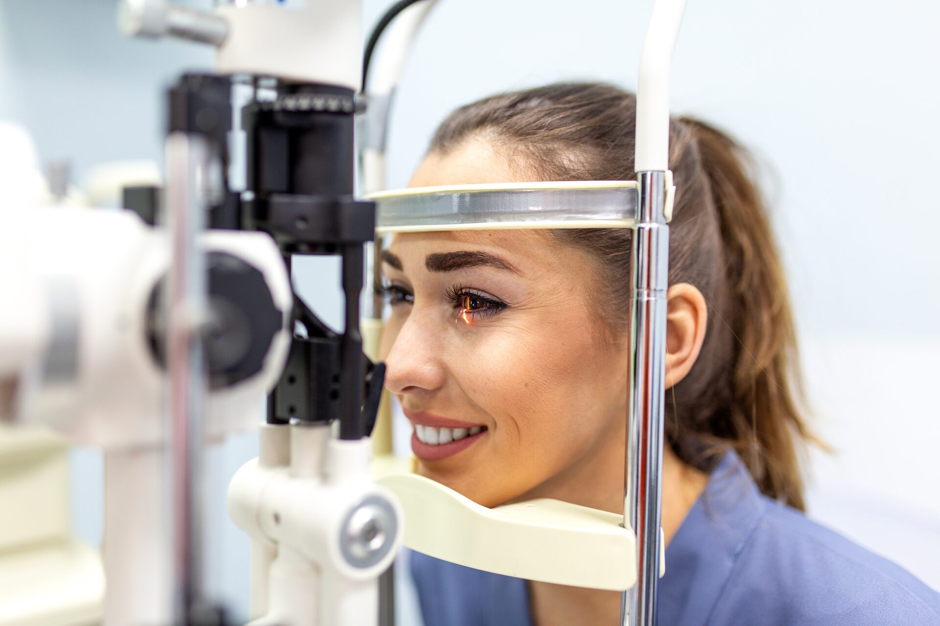
The slit lamp magnifies the ocular surface, cornea, anterior chamber, iris and lens. Tonometry measures intraocular pressure. Corneal thickness, angle anatomy and optic-nerve findings influence interpretation; there is no single “safe” pressure for every nerve.
Dilating drops relax iris muscles and enlarge the pupil. A wider view helps the clinician inspect the lens, vitreous, optic nerve, macula, retinal vessels and more peripheral retina. Dilation supports detection of diabetic retinopathy, AMD, glaucoma-related nerve changes, tears, detachments, inflammation and other problems.
Drops generally take time to work. Light sensitivity and near blur may last several hours, sometimes longer depending on the medication, eye color, age and individual response. Bring sunglasses. Ask whether driving is advisable; people who feel unsafe should arrange transportation.
Dilation can very rarely precipitate angle closure in susceptible eyes. Severe pain, redness, halos, headache, nausea or vomiting after an exam requires immediate contact or emergency care. Clinicians may assess angle risk before dilation when indicated.
More testing is not automatically better. Each test should answer a clinical question, establish a baseline or measure change. Incidental abnormalities need interpretation rather than alarm.
Ask what was normal, what was abnormal, what diagnosis is being considered, what change should trigger contact and when the next visit is due. If monitoring is recommended, understand which measurement is being trended.
Routine intervals do not apply to sudden symptoms. Seek urgent evaluation for a new curtain or shadow, flashes with many floaters, sudden major blur, severe pain, painful red photophobic eye, trauma, chemical exposure or postoperative pain and decreased vision. Call emergency services for visual change with facial droop, weakness, numbness, speech difficulty, severe imbalance or confusion.
Transient symptoms can still be serious. Review When Vision Changes Cannot Wait and act according to the current pattern rather than waiting for a scheduled exam.
NRT at Netra Eye Institute is a supportive functional service, not a screening substitute. A patient needs diagnosis and medical stabilization before persistent symptoms are assigned to rehabilitation.
When eye health is stable, assessment may examine accommodation, binocular coordination, scanning, eye–head control, visual attention, motion sensitivity, field awareness and endurance. NRT may be coordinated with updated optical correction, ophthalmic care, low-vision services, occupational therapy, physical therapy or neurology.
Goals should reflect daily function, such as reading for a defined time, finding targets in a field-loss area or tolerating a visually complex environment. NRT does not lower glaucoma pressure, remove cataract, treat retinal leakage or replace anti-VEGF injections and surgery.
Learn about Neuro-Visual Rehabilitation Therapy, Netra Eye Institute’s approach or contact the Institute after appropriate medical evaluation.
Yes at an interval appropriate to risk. Glaucoma, diabetic retinopathy and early AMD may be asymptomatic. Clear acuity does not prove a healthy optic nerve or peripheral retina.
No. Photography documents a defined field and can be valuable, but dilation permits a broader dynamic examination. Whether imaging can answer the immediate question depends on condition and image quality.
Routine diagnostic dilation is generally safe. Temporary near blur and light sensitivity are expected. Severe pain, nausea, halos or persistent concerning symptoms are not expected and require contact.
Often several hours, but duration varies. Reading and close work are usually affected more than distance. Follow the clinic’s driving advice and your own functional safety.
Optometrists provide comprehensive eye care and manage many conditions; ophthalmologists are physicians who provide medical and surgical eye care. Subspecialists focus on retina, glaucoma, cornea, neuro-ophthalmology and other areas. The right entry point depends on urgency and known disease, with referral when needed.
Aging predictably changes near focus, light needs and adaptation, but vision loss should never be dismissed as “just age.” Cataract, glaucoma, AMD, diabetic retinopathy and retinal disease require detection and, when indicated, treatment.
Use age-based recommendations as a starting point and let personal risk determine the actual interval. A comprehensive dilated examination evaluates far more than a glasses prescription, while new warning signs require immediate care regardless of the calendar. After disease is diagnosed and stable, NRT may support selected functional difficulties without replacing medical care.
Medical Disclaimer: This article provides general education and is not medical advice, diagnosis or a personal examination schedule. Follow the interval recommended by your eye-care and medical clinicians. Seek urgent care for sudden loss, a curtain or shadow, new flashes and floaters, severe pain, chemical injury, trauma or visual symptoms with neurologic changes. NRT must not delay established medical, surgical or emergency treatment.



