Search conditions, treatments, research reviews and articles. Press Enter to see all results, Esc to close.
Search conditions, treatments, research reviews and articles. Press Enter to see all results, Esc to close.
Blog
October 31, 2026
Papilledema is swelling of the optic disc, where the optic nerve leaves the eye, caused by raised pressure inside the skull. It is a warning sign that needs medical assessment the same day, even if you see clearly and feel well. Doctors look for the cause with an urgent brain scan, a blood pressure check and often a spinal tap (lumbar puncture) [1,3,5].
This article is general education, not a diagnosis. If an eye doctor has just told you that your optic nerves look swollen, follow their referral today. Do not wait for a routine appointment.
Key takeaways
In this article
Call 911 or go to the nearest emergency department now if you have, or may have, swollen optic discs and any of these:
If you have none of these but an eye exam found swollen optic discs, you still need same-day assessment. Ask the eye doctor exactly where to go and who is expecting you. Don't drive yourself if your vision is affected. If you can, note when each symptom started [11].
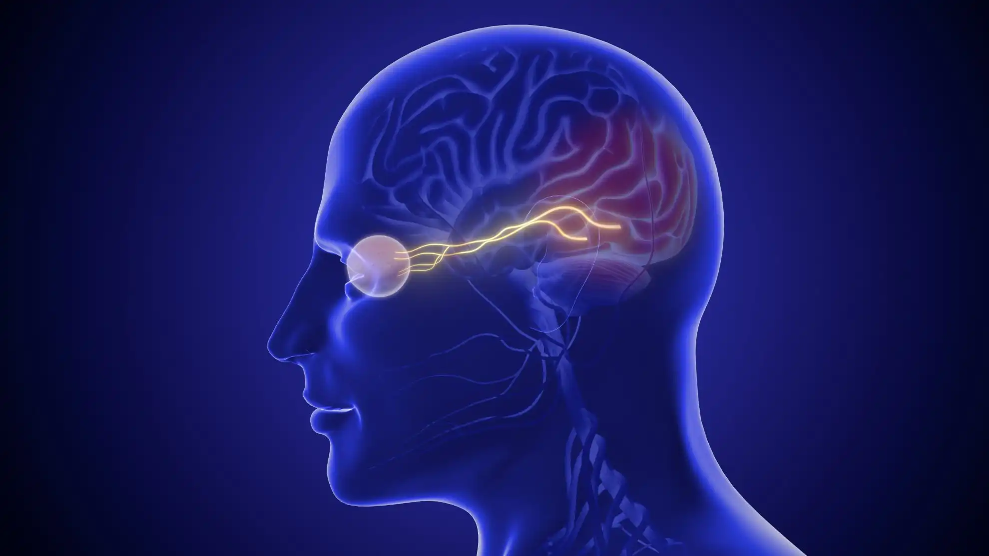
Papilledema is swelling of the optic discs caused by high pressure inside the skull (raised intracranial pressure). The optic disc is where nerve fibers from the retina gather to form the optic nerve. An eye doctor sees the swelling during an exam. It means something is raising the pressure around the brain, so the cause must be found quickly [1,5].
The optic nerve is wrapped in the same layers that cover the brain. The fluid around the brain, called cerebrospinal fluid (CSF), also surrounds the nerve. When that fluid pressure rises, it is passed along to the back of the eye. The pressure slows the normal flow of material inside the nerve fibers, and the fibers swell where they leave the eye [5].

"Optic disc swelling" is the broader term. Doctors use "papilledema" only when raised skull pressure is the cause. Swelling from other causes, such as optic neuritis or ischemic optic neuropathy, is called optic disc edema instead [1].
Papilledema is also different from high eye pressure. The puff or probe test at an eye exam measures pressure inside the eyeball, not inside the skull. A normal eye pressure reading does not rule out papilledema.
Most symptoms come from the high pressure itself. They include headache, brief graying of vision that lasts seconds, and a whooshing sound in the ears that beats with your pulse. Nausea and blurred or double vision can also occur. Sharp central vision is usually normal at first [1,2,5].
The brief dimming spells are called transient visual obscurations. They usually last seconds and often happen when you stand up after bending or lying down [2,5]. Not everyone has them, so their absence does not rule papilledema out.
The pulsing ear sound is called pulsatile tinnitus [2]. High pressure can also affect the sixth cranial nerve, which turns the eye outward. That causes double vision with the two images side by side [6,8].
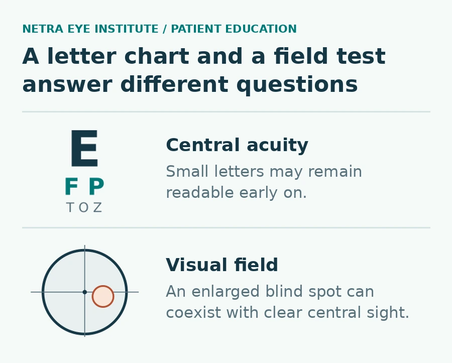
Reading the eye chart tests only the center of your vision. Papilledema first enlarges the natural blind spot, and early vision loss tends to start at the edges [1,6]. People may not notice it until late. For this reason, the eye chart is not sensitive enough to warn of coming vision loss [6].
A visual field test maps how well you see small lights across your side vision while you look straight ahead. It can pick up changes the chart misses. Keep your eye on the center target and don't search for the lights.
Anything that raises pressure inside the skull can cause papilledema. Causes include a brain tumor, abscess or bleed, and blocked flow of brain fluid (hydrocephalus). Others are meningitis or encephalitis, a blood clot in the brain's large veins (venous sinus thrombosis), and head injury. The most common cause is idiopathic intracranial hypertension (IIH) [1,5].
IIH means high brain pressure with no tumor or other cause found. It is most common in women of childbearing age and is strongly linked to obesity [2,6]. It can still affect anyone, so the full workup is needed every time. The diagnosis is made only after other causes have been ruled out [7].
Some medicines are linked to raised brain pressure. Examples include tetracycline antibiotics, high doses of vitamin A, isotretinoin, lithium, and starting or stopping steroids [6,7]. Tell your doctors about every medicine and supplement you take. Do not stop a prescribed medicine without advice.
Very high blood pressure (malignant hypertension) can also swell the optic discs. Strictly speaking, that is not papilledema, but it needs emergency treatment of its own [1,3]. Blood pressure should be measured whenever papilledema is suspected [8]. UK guidelines use a reading of 180 systolic or 120 diastolic or higher as the danger level [3]. The American Heart Association uses the same level: call 911 if your blood pressure is higher than 180/120 and you have symptoms such as a change in vision [17].
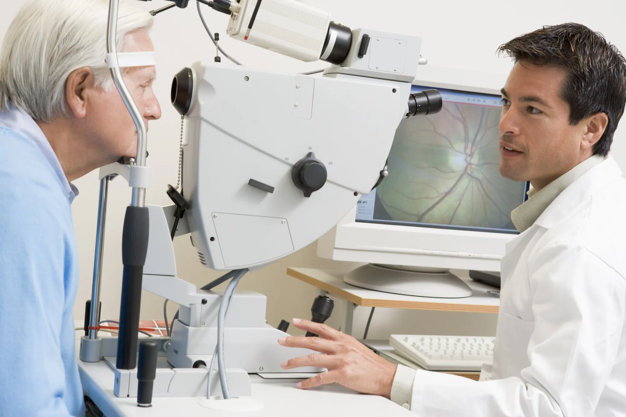
On an eye exam, the optic disc looks raised, swollen and redder than usual. The small blood vessels on its surface can be hidden. Nearby veins can look swollen and twisty, and there may be small bleeds around the disc. You cannot see these changes yourself, and they do not show the cause [1,5].
Doctors grade the swelling from mild to severe, often with the Frisén scale, which runs from 0 (normal) to 5 (most severe) [5]. Photos and OCT scans create a record to compare over time [3]. OCT (optical coherence tomography) is a light-based scan that measures the thickness of the nerve fiber layer [1].
Testing has two goals: confirm that the swelling is real, and find what is raising the pressure. Expect a dilated eye exam, a visual field test and a blood pressure check. Next comes an urgent brain MRI or CT that includes the brain's veins. A lumbar puncture then usually measures the fluid pressure [1,3].
| Test | What it shows | What it cannot tell you alone |
|---|---|---|
| Dilated eye exam and photos | Disc swelling, its grade, bleeds near the disc | The cause or the pressure number |
| Visual field test | Blind spot size and side-vision loss | Why the field changed |
| OCT scan | Nerve fiber thickness over time | Whether thinning means healing or nerve loss |
| Blood pressure | Dangerously high blood pressure | Pressure inside the skull |
| MRI or CT with venography | Tumors, blocked fluid flow, clots in the brain's veins | The exact brain pressure |
| Lumbar puncture | Opening pressure and signs of infection or inflammation in the fluid | The full picture without the scan and eye findings |
The scan comes before the spinal tap. A lumbar puncture in someone with a brain mass can be dangerous, so imaging checks for this first [1]. UK consensus guidelines advise a brain MRI within 24 hours, or CT if MRI is not available. They also call for a vein scan (MR or CT venography) to rule out a clot [3].
During the lumbar puncture, the doctor measures the opening pressure. This is the fluid pressure before any fluid is removed, recorded in centimeters of water. In adults, a reading above 25 cm is the usual cutoff for IIH. It is read together with the scan, the eye findings and the fluid tests, not on its own [3,6].
Scans can show signs that fit high pressure, but they cannot replace the lumbar puncture [2]. If you have old eye photos or earlier scans, bring them or a link to them. Don't delay an urgent visit to collect records.
Sometimes. An optic disc can look swollen when it is not. This is called pseudopapilledema. Common causes are optic disc drusen (small deposits in the nerve head) and disc shapes some people are born with, such as a tilted disc. These are often harmless, but only an eye specialist with the right tests can tell them apart from true swelling [8,9].
Drusen are usually found by chance during an eye exam, and most people have no symptoms. Up to a quarter have side-vision defects, so drusen are checked from time to time [9]. Tests that help tell drusen from swelling include an ultrasound of the eye (B-scan), OCT, and autofluorescence photos, in which drusen glow [1,9]. Old eye photos that show the same look years ago also help.
Having drusen does not protect you from other problems. A new headache or vision change still needs prompt care. And when only one disc is swollen, doctors look harder for a cause in that eye, because true papilledema in one eye is rare [8].
| Feature | Papilledema | Pseudopapilledema (such as drusen) | Other optic disc swelling |
|---|---|---|---|
| What is happening | True swelling from high pressure in the skull | The disc only looks swollen | True swelling from inflammation, poor blood flow or very high blood pressure |
| Urgency | Same-day assessment | Needs specialist confirmation | Prompt assessment; very high blood pressure is an emergency |
| Key tests | Brain MRI or CT with venography, lumbar puncture, visual fields | Eye ultrasound, OCT, autofluorescence, old photos | Eye exam, visual fields, blood pressure, and other tests based on the cause |
Treatment targets the cause of the high pressure. A tumor, blood clot, infection or blocked fluid flow is treated by the right medical team. For IIH, the main treatments are weight loss, for people with overweight or obesity, and medicine such as acetazolamide. Surgery is used when vision gets worse despite treatment [1,2,3].
Weight loss is the only treatment shown to change the course of typical IIH [3]. A common first goal is losing about 6 to 10 percent of body weight [2]. Ask your team for real support with this, not just advice.
Acetazolamide is the most common medicine for IIH [2]. In the IIH Treatment Trial, 165 people with IIH and mild vision loss followed a low-salt weight-loss diet for six months. They also took either acetazolamide or a placebo. The acetazolamide group had a modest extra gain in visual field scores, less disc swelling and better quality of life [4]. The trial included only people with mild vision loss. It does not mean someone whose sight is failing fast should wait for tablets to work [3,4].
Common side effects include tingling, tiredness, taste changes and nausea. Kidney stones are rare [3]. Don't change the dose on your own. Topiramate is another option [1,2]. If you are pregnant or planning pregnancy, talk to your team early. Guidelines say safe advice on acetazolamide in pregnancy is hard to give [3].
Fewer than 1 in 10 people with IIH need surgery [2]. Options include a shunt that drains brain fluid and optic nerve sheath fenestration, a small opening in the sheath around the optic nerve. Some centers also place a stent in a narrowed brain vein [1,2]. The choice depends on the cause, how fast vision is changing and local expertise [3].
Headache can continue even after the swelling settles. Taking simple pain relievers on more than 15 days a month can itself keep headaches going [3]. Ask for a written headache plan and a separate vision plan, so a good headache week never cancels a field test.
Once the pressure is brought down, well-developed papilledema usually settles over about 1.5 to 2.5 months [5]. The outlook for sight depends on how high the pressure was and for how long. Vision already lost to nerve damage usually does not return, even after the pressure is normal again [6].
If high pressure goes on untreated, the nerve can waste away (optic atrophy), causing permanent vision loss [1,5]. Netra's guide to optic nerve damage explains why the cause and location of an injury matter.
A falling OCT number needs careful reading. As swelling goes down, the nerve fiber layer gets thinner. That can mean healing, or it can mean nerve fibers have been lost. Doctors compare OCT with visual fields and with scans of the retina's ganglion cell layer to tell the difference [10].
Formal visual field testing is used to document and follow any vision loss [8]. If fields or swelling get worse, your next visit should be brought forward [3]. Keep dated copies of field tests, scan reports and opening pressure results. Ask who reviews your fields and who to call if your vision changes.
Not as a treatment. Netra Restoration Therapy (NRT) has not been tested in controlled trials for papilledema or IIH. It does not lower brain pressure, treat a blood clot or tumor, or rescue vision during a crisis. Swollen optic discs need urgent medical care first, and NRT should never delay it [15,16].
Netra Restoration Therapy is an integrative program based on traditional Chinese medicine and Ayurvedic principles, including acupuncture. Netra uses it alongside the diagnosis, monitoring and treatment from your eye doctors, never instead of them [15].
NRT may come up only later. That means after the cause is found and treated, and once vision and pressure are stable. A person with lasting side-vision loss may then ask about complementary support for daily function. Bring your diagnosis, field tests, OCT reports and your specialist's follow-up plan. Any goal should be measurable, such as time to find items on a cluttered counter.
Netra explains how it reviews outcomes by diagnosis on its NRT effectiveness page [16]. A better headache or feeling more relaxed does not show that your field or brain pressure has changed. Practical help from low vision rehabilitation may also be useful.
Pause any complementary care and call your medical team if dimming spells return or double vision starts. Do the same for any new nerve symptom or a worse field test.
Yes. Newly found papilledema is treated as a medical emergency because the cause can be serious and vision can be lost [5]. You need same-day assessment with urgent brain imaging, even if you feel well [1,3]. Call 911 for sudden vision loss, a sudden severe headache, weakness or confusion [11].
The swelling usually goes away once the cause is treated and the pressure comes down [5]. IIH can often be controlled with weight loss, medicine and sometimes surgery [2]. Vision already lost to nerve damage usually does not come back, which is why fast treatment and follow-up matter [6].
It can. If high pressure is not treated, the optic nerve can be permanently damaged [1]. For IIH, NANOS notes that relatively few people go blind, and most recover when treated early. Severe untreated cases, though, carry a high risk of lasting vision loss [2].
Yes. Headache is the most common symptom of IIH, but not everyone has it [2]. Sharp central vision is also usual early on [1]. That is why swollen discs found at a routine eye exam still need same-day follow-up, even if you feel fine.
Almost always both, although one eye may look worse than the other [1,8]. Papilledema in only one eye is rare [8]. When just one disc is swollen, doctors also check that eye for other problems, such as optic neuritis or poor blood flow to the nerve [1].
Optic disc edema means any swelling of the optic disc. Papilledema is the name used only when raised pressure inside the skull is the cause [1]. Inflammation, poor blood flow and very high blood pressure can also swell the disc, but those are not called papilledema [1].
No. Eye pressure is the pressure inside the eyeball, measured at a routine eye exam. Papilledema comes from high pressure inside the skull, which is measured during a lumbar puncture [3]. A normal eye pressure reading does not rule out papilledema.
Sources checked October 31, 2026.
This article provides general education and does not replace an individual eye examination, diagnosis or treatment plan. If you have been told your optic discs look swollen, get medical assessment the same day, and call 911 for sudden vision loss, a sudden severe headache, weakness or confusion.