
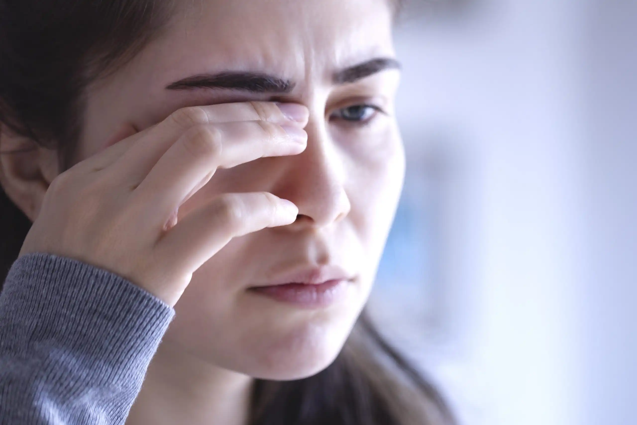
Corneal neuralgia is not simply dryness or irritation. It is a pain-centered disorder in which corneal nerve injury, ocular surface stress, inflammation, and altered trigeminal processing may generate burning, aching, stabbing pain, photoallodynia, or pain from ordinary airflow. Netra Restoration Therapy is designed as an adjunctive approach that supports the biological terrain influencing ocular surface comfort, peripheral nerve health, and nervous-system regulation.
1. NRT - A Comprehensive Therapy Designed to Support the Key Underlying Drivers of Corneal Neuralgia
Corneal neuralgia, commonly discussed as neuropathic corneal pain, involves abnormal activity in sensory pathways connecting the cornea and trigeminal nervous system. Patients may describe burning, stabbing, aching, pressure, electric sensations, pain from wind, or severe light sensitivity. Symptoms may be constant or episodic and disproportionate to routine ocular surface findings.
The word neuralgia emphasizes pain. Corneal neuropathy is broader and may produce reduced sensation, abnormal sensation, pain, or mixed findings. Corneal neuralgia is the painful expression of abnormal corneal or trigeminal signaling. It also differs from neurotrophic keratopathy, where reduced sensation and impaired healing can lead to epithelial breakdown. Overlap is possible, but the terms are not interchangeable.
The cornea is exceptionally well innervated. Its sensory nerves detect touch, temperature, evaporation, chemical change, and potential injury. They also participate in reflex blinking, tear secretion, epithelial renewal, wound healing, and immune communication. When these nerves are injured or repeatedly provoked, their firing thresholds may fall. Ordinary sensations such as airflow, light, tear evaporation, or blinking can then become painful. If abnormal input continues, pain processing in the trigeminal brainstem and higher nervous system may also become amplified.
Netra Restoration Therapy, or NRT, is an integrative platform designed to support ocular surface comfort, corneal nerve resilience, inflammatory balance, and nervous-system regulation. It is not a cure, and the combined protocol has not been established for corneal neuralgia in controlled trials. NRT is intended to complement evaluation and treatment by qualified professionals, including other specialties when broader pain or systemic involvement is suspected.
For patients with stable chronic symptoms, NRT is designed to consider several interacting domains:
- Ocular surface and epithelial barrier health
- Tear-film stability and evaporative stress
- Corneal nerve irritation, resilience, and repair signaling
- Peripheral sensitization at corneal nerve endings
- Central or nociplastic pain amplification
- Neurogenic inflammation and immune signaling
- Oxidative and mitochondrial stress
- Autonomic nervous-system balance
- Sleep, migraine, stress physiology, and pain thresholds
- Metabolic, autoimmune, nutritional, and digestive contributors
- Functional limitations and quality-of-life burden
Corneal neuralgia is rarely explained by one factor. Surgery, infection, contact lens injury, dry eye, or inflammation may initiate symptoms, while tear instability, peripheral firing, central sensitization, migraine, poor sleep, or systemic pain vulnerability may sustain them. NRT uses a multi-factorial model after urgent or sight-threatening causes are excluded.
2. Why Support for Corneal Neuralgia Should Be Multi-Factorial
Corneal neuralgia should be approached as a multi-factorial pain condition because the corneal sensory system is inseparable from the tear film, epithelium, immune system, trigeminal pathway, and the patient’s broader pain physiology. Lubrication alone may be insufficient when nerve endings are hypersensitive, while focusing only on central pain can overlook surface disease that continues to drive peripheral input.
Corneal Nerve Injury and Ectopic Firing
Corneal nerve injury may follow refractive surgery, cataract surgery, corneal infection, trauma, chronic ocular surface disease, chemical exposure, contact lens stress, diabetes, systemic neuropathy, or inflammatory disease. During repair, regenerating nerve endings may develop altered ion-channel expression, abnormal excitability, disorganized branching, or neuroma-like changes. Injured fibers can then fire spontaneously or respond excessively to weak stimuli.
The result may be burning at rest, sharp pain with blinking, pain from a fan, or intolerance of ordinary light. These symptoms are biologically consistent with an altered sensory threshold.
Tear-Film Instability and Repeated Peripheral Provocation
The tear film separates corneal nerve endings from air, friction, osmotic change, and irritants. Rapid breakup repeatedly stresses the epithelium and nerves, while nerve damage may reduce reflex tearing and blink quality. Patients can therefore have both dry eye disease and corneal neuralgia; surface and pain-system findings should be assessed together.
Peripheral Sensitization
Peripheral sensitization occurs when nociceptors at or near the corneal surface become easier to activate. Inflammatory mediators, epithelial injury, hyperosmolar stress, and nerve damage can alter receptors and ion channels involved in sensing temperature, chemicals, and mechanical stimuli. Harmless airflow may be experienced as burning; normal light may trigger pain through trigeminal pathways; and mild dryness may feel severe.
A clinician may use a topical anesthetic challenge as one piece of the evaluation. Substantial temporary relief after anesthetic suggests a strong peripheral contribution. Incomplete or absent relief may suggest central amplification, but the test is not a stand-alone diagnostic rule and mixed pain is common.
Central Sensitization and Nociplastic Features
Persistent peripheral input can change how pain is processed in the trigeminal brainstem and higher neural networks. Central sensitization can involve increased excitatory signaling, reduced inhibition, glial activation, and expansion of the response to normally non-painful stimuli. Pain may spread beyond the ocular surface or coexist with photophobia, migraine, headache, facial pain, sound sensitivity, fibromyalgia, or generalized sensory hypersensitivity.
When central mechanisms are prominent, surface improvement may not fully resolve pain. Care may need to include neuropathic-pain or migraine expertise, sleep restoration, graded rehabilitation, and support for coping with persistent pain.
Neuroinflammation and Neurogenic Inflammation
Stressed epithelial cells, immune cells, and injured nerves can release cytokines, chemokines, prostaglandins, ATP-related signals, substance P, and calcitonin gene-related peptide. These mediators may lower nociceptor thresholds and sustain abnormal signaling. Sensory nerves can also influence local blood vessels and immune cells, creating a feedback loop in which inflammation sensitizes nerves and activated nerves promote further inflammation.
NRT frames inflammatory balance as one domain of support. It should not be used to delay medical treatment for infection, uveitis, scleritis, keratitis, corneal ulceration, or another inflammatory disorder that requires prompt diagnosis.
Neurotrophic Signaling and Repair Biology
Corneal nerves and epithelial cells support one another through neurotrophins, neuropeptides, and growth factors. Nerve growth factor, or NGF, has important roles in corneal epithelial healing, nerve survival, sensitivity, and ocular surface homeostasis. Other mediators, including brain-derived neurotrophic factor, may participate in sensory plasticity, although their exact clinical role in corneal neuralgia is still being defined.
The relevance of a biological pathway does not prove that a supplement, herb, acupuncture protocol, or integrative program changes it in patients. NRT uses nerve-epithelium science as a rationale for supporting tissue health, not as proof of cure or nerve regeneration.
Oxidative, Mitochondrial, and Metabolic Stress
Peripheral nerves require sustained energy to maintain membrane gradients, transport cellular materials, and repair injury. Oxidative and mitochondrial stress may be increased by chronic inflammation, diabetes, nutritional insufficiency, poor sleep, metabolic disease, or environmental exposure. These factors may weaken the tissue environment in which nerves and epithelial cells attempt to recover.
NRT considers metabolic and antioxidant capacity as supportive terrain, not as a stand-alone reversal of established sensitization.
Autonomic Regulation, Sleep, and Stress Physiology
Autonomic activity influences lacrimal secretion, blink patterns, blood flow, immune tone, arousal, and pain modulation. Poor sleep can lower pain thresholds and worsen migraine, fatigue, mood, and coping. Stress may intensify symptoms through physiological arousal without being the original cause of the disorder.
A systems-based plan considers sleep, autonomic arousal, activity tolerance, and daily triggers while ocular and medical causes are addressed.
3. Key Biological and Clinical Mechanisms in Corneal Neuralgia
The Corneal-Trigeminal Pain Pathway
Corneal sensory fibers travel through the ophthalmic division of the trigeminal nerve. Signals enter brainstem regions that integrate ocular sensation with facial pain, blinking, tearing, and light-related responses. This anatomy helps explain why corneal neuralgia may be felt as pain in the eye, brow, temple, face, or head and why migraine and photophobia frequently overlap with ocular surface pain.
Corneal neuralgia may be predominantly peripheral, predominantly central, or mixed. These categories describe the suspected location of pain generation; they are not always fixed. A patient can begin with a peripheral injury and later develop central amplification, while persistent ocular surface triggers continue to supply peripheral input.
Allodynia, Hyperalgesia, and Photoallodynia
Allodynia means pain caused by a stimulus that is normally not painful. In corneal neuralgia, examples include pain from air movement, gentle touch around the eye, blinking, or ordinary indoor light. Hyperalgesia means an exaggerated response to a stimulus that is already painful. Photoallodynia describes pain provoked by light rather than simple visual discomfort.
These symptom qualities are clinically useful because ordinary dry eye questionnaires may not fully capture electric, burning, evoked, or radiating pain. Neuropathic-pain questionnaires adapted for the eye, such as the NPSI-Eye, can help characterize the pain phenotype, but no questionnaire establishes the diagnosis by itself.
Symptom-Sign Discordance
Some patients have severe pain with minimal staining or redness. This discordance can occur because a slit-lamp examination shows tissue structure and surface disease but does not directly measure all abnormal nerve activity or central pain processing. At the same time, normal-looking findings must not become a shortcut to diagnosis. Infection, inflammation, exposure, recurrent erosion, intraocular disease, headache disorders, and referred pain should be considered according to the clinical presentation.
The phrase “pain without stain” should never be used dismissively. Limited staining does not invalidate pain; it signals the need for a broader differential diagnosis.
Corneal Sensitivity and Esthesiometry
Corneal sensitivity can be assessed qualitatively with a cotton wisp or quantitatively with esthesiometers. Patients with corneal neuralgia may show increased, reduced, asymmetric, or apparently normal sensitivity depending on disease stage and testing method. Hyperesthesia may support peripheral sensitization, while reduced sensation may indicate nerve injury or overlap with neurotrophic dysfunction.
No single sensitivity value confirms or excludes corneal neuralgia; results require clinical context.
In Vivo Confocal Microscopy
In vivo confocal microscopy, or IVCM, can visualize the corneal subbasal nerve plexus. Studies in neuropathic corneal pain have reported changes such as lower nerve density, increased tortuosity or reflectivity, beading, abnormal branching, and microneuroma-like structures. These findings may support a nerve injury phenotype and can be useful in specialized centers.
IVCM is not definitive or universally available. Similar abnormalities can occur in dry eye, diabetes, and after surgery. Microneuromas remain under investigation, and an equivocal scan does not exclude centrally maintained pain.
The Topical Anesthetic Challenge
A preservative-free topical anesthetic may temporarily silence peripheral corneal input. Marked relief suggests that ongoing peripheral signals contribute substantially to the pain. Persistent pain after the surface is anesthetized suggests central or extra-corneal mechanisms, although incomplete anesthesia, mixed pain, and expectation effects limit interpretation.
Topical anesthetics are clinician-administered diagnostic tools, not medicines for unsupervised repeated use, which can damage the corneal epithelium.
Ocular Surface Inflammation and Tear Hyperosmolarity
Dry eye disease, blepharitis, meibomian gland dysfunction, allergy, preservative toxicity, and incomplete blinking can maintain inflammatory or osmotic stress at the nerve endings. Treating these contributors is important even when central sensitization is present because reducing peripheral input may lower the continuing load on the pain system.
Neurotrophic Keratopathy Is Different
Neurotrophic keratopathy is characterized by reduced corneal innervation and impaired epithelial healing, often with reduced sensation. Corneal neuralgia is characterized by pain and sensory amplification. A person can paradoxically have reduced measured sensation in one area and painful dysesthesia in another, but the management priorities differ. Persistent epithelial defects, ulceration, or stromal thinning require urgent corneal care and should not be managed as neuralgia alone.
Systemic Pain, Migraine, and Small-Fiber Neuropathy
Corneal neuralgia may coexist with migraine, fibromyalgia, post-herpetic neuralgia, diabetes, autoimmune disease, anxiety, depression, sleep disturbance, and small-fiber neuropathy. These associations do not prove that every patient has systemic neuropathy. They do support asking whether ocular pain is part of a broader sensory phenotype.
Symptoms beyond the eye may warrant neurological, rheumatological, metabolic, or pain evaluation.
4. What Is Netra Restoration Therapy for Corneal Neuralgia?
Netra Restoration Therapy is an individualized integrative program designed to support ocular health through multiple interacting biological and functional domains. For corneal neuralgia, NRT focuses on the ocular surface, corneal nerve environment, inflammatory balance, tear-film stability, autonomic regulation, lifestyle contributors, and the patient’s broader recovery terrain.
Depending on clinical appropriateness, NRT may incorporate acupuncture-based care, principles from Traditional Chinese Medicine and Ayurveda, nutrition and botanical review, lifestyle guidance, sleep and stress support, digestive and metabolic assessment, and coordination with conventional eye-care recommendations. Any botanical or nutritional plan should be reviewed for interactions, contraindications, pregnancy considerations, kidney or liver disease, anticoagulant use, and other medical risks.
NRT does not diagnose corneal neuralgia solely from symptoms or replace established care. Depending on the phenotype, conventional management may address ocular surface disease, inflammation, migraine, or neuropathic pain and may use protective lenses, biologic tear products, medication, procedures, or multidisciplinary rehabilitation. Patients should not stop prescribed treatment without consulting the responsible clinician.
For a stable patient being considered for adjunctive support, the NRT framework asks:
- Have infection, corneal ulceration, uveitis, scleritis, glaucoma-related emergencies, and other urgent causes been excluded?
- Is there active dry eye, blepharitis, allergy, exposure, epithelial disease, or preservative toxicity?
- Did symptoms begin after surgery, infection, trauma, contact lens use, or an inflammatory flare?
- Are pain qualities neuropathic, such as burning, electric pain, allodynia, hyperalgesia, or photoallodynia?
- Does the pattern suggest peripheral, central, or mixed pain?
- Are migraine, facial pain, small-fiber neuropathy, autoimmune disease, diabetes, or systemic pain present?
- Are sleep disruption, fear of triggers, inactivity, or stress physiology amplifying disability?
- Which supportive strategies can be added without delaying or interfering with medical care?
This framework is intended to individualize supportive care. It is not a diagnostic algorithm or a promise of response.
5. How NRT Supports the Biological Terrain in Corneal Neuralgia
Supporting the Ocular Surface-Nerve Interface
The first objective is to reduce unnecessary provocation of nerve endings. NRT considers hydration, blinking, screens, airflow, sleep, nutrition, lid and tear-film factors, and adherence to established ocular surface care. Worsening findings require medical reassessment.
Supporting Corneal Nerve Resilience and Neurotrophic Terrain
NRT is designed to support the tissue conditions associated with nerve and epithelial health: adequate nutrients, metabolic stability, balanced inflammation, restorative sleep, and reduced ongoing surface stress. NGF and other growth factors are scientifically relevant to corneal homeostasis, but NRT should not be marketed as directly increasing NGF or regenerating nerves unless that specific claim has been demonstrated in appropriately designed human studies.
Biologic tear preparations have been studied in neuropathic corneal pain, but their use and monitoring belong under licensed eye-care supervision. NRT can complement, not replace, such care.
Supporting Inflammatory Balance
NRT may use dietary, lifestyle, acupuncture, or botanical strategies to support inflammatory balance, but direct evidence for an NRT protocol in corneal neuralgia is not yet available.
Traditional Chinese Medicine may describe patterns using terms such as Yin deficiency, Blood deficiency, Heat, Qi stagnation, Blood stasis, or Liver Wind. Ayurveda may use concepts such as Vata, Pitta, Rakta Dhatu, Majja Dhatu, and Ojas. These are traditional clinical frameworks. They can guide individualized traditional practice but are not exact translations of peripheral sensitization, cytokine activity, small-fiber neuropathy, or central pain processing.
Supporting Autonomic Balance and Pain Modulation
Acupuncture has been studied more extensively in dry eye disease than in confirmed corneal neuralgia. Some clinical studies report improvements in ocular discomfort, tear-film measures, corneal sensitivity, or mood-related outcomes, and preclinical research suggests possible effects on inflammatory and sensory signaling. The evidence is heterogeneous, and it does not establish acupuncture as a cure for neuropathic corneal pain.
Within NRT, acupuncture is one component intended to support autonomic regulation and function. Central sensitization may still require medication, migraine treatment, pain rehabilitation, or specialist care.
Supporting Mitochondrial and Metabolic Health
Because nerve function is energy-dependent, NRT reviews factors that can impair cellular resilience, including poorly controlled diabetes, nutritional deficiency, sleep loss, systemic inflammation, and metabolic instability. Correcting a documented deficiency or improving metabolic health may support overall nerve health, but it should not be presented as a proven stand-alone treatment for corneal neuralgia.
Supporting Sleep and Functional Recovery
Chronic ocular pain can disrupt reading, computer work, driving, social activity, and sleep. In turn, insomnia and inactivity can increase pain sensitivity and reduce resilience. NRT encourages gradual restoration of sleep routines, appropriate physical activity, pacing, and exposure management in coordination with the patient’s medical team.
The aim is not forced exposure, but avoidance of progressive sensory restriction, deconditioning, and disability.
Considering the Gut-Eye and Immune-Metabolic Axis
The gut-eye axis is an emerging area of research, especially in dry eye disease and autoimmune conditions such as Sjogren syndrome. Microbiome composition, intestinal barrier function, microbial metabolites, and systemic immune signaling may influence the lacrimal gland and ocular surface. Direct evidence that microbiome modification treats corneal neuralgia is not established.
Digestive, nutritional, autoimmune, and metabolic factors may be considered as individualized supportive terrain, not as universal causes of corneal pain.
Supporting Systems-Level Botanical Care Safely
Botanical formulas contain multiple bioactive compounds and are often used in traditional systems to address several symptom and constitutional patterns at once. Laboratory and network-pharmacology findings can help generate hypotheses, but they do not prove clinical effectiveness for corneal neuralgia. Product quality, dosing, interactions, and organ function matter.
Herbal care should be prescribed by a trained clinician who reviews medications and medical history. Non-sterile herbal preparations must never be placed in the eye.
Supporting the Whole Person Without Invalidating the Pain
Persistent corneal pain can produce anxiety, depressed mood, hypervigilance, and loss of confidence. These are understandable consequences and possible amplifiers of chronic pain, not proof that the disorder is imaginary. A compassionate plan validates the biological reality of the symptoms while addressing the patient’s emotional, social, and functional needs.
The aim of NRT is to provide coordinated adjunctive support across the ocular surface, nervous system, sleep, nutrition, inflammation, metabolism, and daily function. Outcomes vary, and improvement cannot be guaranteed.
Frequently Asked Questions on Corneal Neuralgia
Corneal neuralgia is persistent or recurrent pain arising from abnormal signaling in corneal sensory nerves and the trigeminal pain pathway. It is commonly called neuropathic corneal pain. Symptoms may include burning, stabbing, aching, electric sensations, foreign-body sensation, wind sensitivity, and painful light sensitivity.
Not exactly. Corneal neuropathy is the broader category of corneal nerve injury or dysfunction and may cause reduced sensation, abnormal sensation, pain, or impaired protective reflexes. Corneal neuralgia refers specifically to the painful phenotype. A patient with neuralgia may have corneal neuropathy, but not every corneal neuropathy causes pain.
No. Neurotrophic keratopathy is primarily characterized by reduced sensation and impaired epithelial healing, sometimes with persistent defects or ulceration. Corneal neuralgia is characterized by abnormal pain. Overlap is possible, which is why corneal sensation and epithelial integrity should be assessed.
Dry eye disease involves tear-film and ocular surface dysfunction. Corneal neuralgia involves abnormal pain processing. Many patients have both. Neuropathic features are more likely when there is burning or electric pain, allodynia to wind or light, marked symptom-sign mismatch, pain extending beyond the eye, or limited response to surface treatment.
Routine examination can exclude many dangerous causes and show surface damage, but it does not directly measure all nerve firing or central sensitization. Abnormal corneal or trigeminal signaling may cause severe pain despite limited visible findings. A normal-looking surface should lead to a broader evaluation, not dismissal of the symptoms.
Reported triggers include refractive or cataract surgery, chronic dry eye disease, infection such as herpes zoster or herpes simplex, trauma, contact lens injury, chemical exposure, diabetes, autoimmune disease, and systemic neuropathy. Some cases have multiple contributors, and in some patients no single initiating event is identified.
Allodynia is pain from a normally non-painful stimulus, such as indoor light or gentle airflow. Hyperalgesia is an exaggerated response to a painful stimulus. Both can occur when peripheral or central sensory pathways become sensitized.
Photoallodynia is pain triggered by light. It may overlap with migraine-related photophobia and may involve corneal input, trigeminal pathways, and central light-pain networks. Severe or new light sensitivity also requires evaluation for inflammation, infection, abrasion, or other ocular disease.
There is no single definitive office test. Diagnosis is clinical and requires exclusion of other causes. Evaluation may include history, slit-lamp examination, staining, tear and lid assessment, corneal sensitivity testing, pain questionnaires, a supervised topical anesthetic challenge, and, in selected centers, in vivo confocal microscopy. Neurological or pain evaluation may be needed.
No. NRT should not be described as a cure or a proven replacement for medical treatment. It is an adjunctive integrative platform designed to support ocular surface health, nerve resilience, inflammatory balance, autonomic regulation, sleep, metabolism, and whole-person recovery.
Direct clinical evidence in confirmed corneal neuralgia is limited. Studies in dry eye-related ocular pain and preclinical models suggest possible effects on discomfort, ocular surface measures, and pain signaling, but study quality and applicability vary. Acupuncture should be considered complementary and individualized.
No. It is a biological pain disorder. Mood, stress, sleep, and attention can influence any chronic pain condition, but they do not make the pain imaginary. Mental-health or pain-psychology support can be valuable for coping and function while ocular and neurological mechanisms are treated.
Seek urgent evaluation for sudden vision loss, intense or rapidly worsening pain, marked redness, trauma, chemical exposure, discharge, a contact-lens-related painful red eye, suspected infection, severe new light sensitivity, halos with nausea, new neurological symptoms, or a persistent epithelial defect. These findings may indicate conditions other than neuralgia that threaten vision or health.
Patients with a stable diagnosis or suspected neuropathic ocular pain may consider an integrative consultation after appropriate medical evaluation. Suitability depends on the ocular examination, pain phenotype, medications, systemic health, medical team, treatment goals, and ability to participate safely in adjunctive care.
Selected References for Scientific Support
- Watson SL, Le DTM. Corneal neuropathic pain: a review to inform clinical practice. Eye (Lond). 2024. This recent clinical review distinguishes peripheral and central mechanisms and summarizes diagnostic and treatment considerations.
- Dieckmann G, Goyal S, Hamrah P. Neuropathic corneal pain: approaches for management. Ophthalmology. 2017. This review outlines history, examination, topical anesthetic challenge, IVCM, and phenotype-based management.
- Goyal S, Hamrah P. Understanding neuropathic corneal pain: gaps and current therapeutic approaches. Semin Ophthalmol. 2016. This paper reviews corneal nerve injury, sensitization, and gaps in evidence.
- Belmonte C, Nichols JJ, Cox SM, et al. TFOS DEWS II pain and sensation report. Ocul Surf. 2017. This consensus report describes ocular surface sensory physiology and pain mechanisms relevant to dry eye and neuropathic ocular pain.
- Galor A, Moein HR, Lee C, et al. Neuropathic pain and dry eye. Ocul Surf. 2018. This review explains neuropathic features, somatosensory dysfunction, and overlap between dry eye symptoms and chronic pain.
- Galor A, Levitt RC, Felix ER, Martin ER, Sarantopoulos CD. Neuropathic ocular pain: an important yet under-evaluated feature of dry eye. Eye (Lond). 2015. This review discusses dysesthesia, allodynia, hyperalgesia, and sensitization.
- Ross AR, Al-Aqaba MA, Almazmi A, et al. Clinical and in vivo confocal microscopic features of neuropathic corneal pain. Br J Ophthalmol. 2020. This prospective observational study evaluated severe pain with minimal visible signs, topical anesthetic response, esthesiometry, and IVCM findings.
- Moein HR, Akhlaq A, Dieckmann G, et al. Visualization of microneuromas by using in vivo confocal microscopy: an objective biomarker for the diagnosis of neuropathic corneal pain? Ocul Surf. 2020. This study evaluated microneuroma-like structures as a potential IVCM biomarker; further validation remains important.
- Crane AM, Feuer W, Felix ER, et al. Evidence of central sensitisation in those with dry eye symptoms and neuropathic-like ocular pain complaints: incomplete response to topical anaesthesia and generalised heightened sensitivity to evoked pain. Br J Ophthalmol. 2017. This study supports the concept that incomplete anesthetic response and generalized pain sensitivity may identify central contributions.
- Farhangi M, Feuer W, Galor A, et al. Modification of the Neuropathic Pain Symptom Inventory for use in eye pain (NPSI-Eye). Pain. 2019. This study supports a questionnaire for characterizing neuropathic qualities of ocular pain.
- Aggarwal S, Colon C, Kheirkhah A, Hamrah P. Efficacy of autologous serum tears for treatment of neuropathic corneal pain. Ocul Surf. 2019. This small retrospective case-control study associated symptom improvement with changes in corneal nerve parameters; it does not establish efficacy through a randomized trial.
- Aggarwal S, Kheirkhah A, Cavalcanti BM, et al. Autologous serum tears for treatment of photoallodynia in patients with corneal neuropathy: efficacy and evaluation with in vivo confocal microscopy. Ocul Surf. 2015. This retrospective study examined photoallodynia, nerve morphology, and response to autologous serum tears.
- Sacchetti M, Lambiase A. Neurotrophic factors and corneal nerve regeneration. Neural Regen Res. 2017. This review summarizes neurotrophic signaling and corneal nerve-epithelium interactions.
- Labetoulle M, Baudouin C, Calonge M, et al. Role of corneal nerves in ocular surface homeostasis and disease. Acta Ophthalmol. 2019. This review links innervation with epithelial integrity, tearing, healing, and pain.
- Dhaliwal DK, Zhou S, Samudre SS, Lo NJ, Rhee MK. Acupuncture and dry eye: current perspectives. A double-blinded randomized controlled trial and review of the literature. Clin Ophthalmol. 2019. The study does not specifically establish treatment for corneal neuralgia.
- Duan H, Zhou Y, Ma B, et al. Effect of acupuncture treatment on ocular pain, mental state and ocular surface characteristics of patients with dry eye disease: a non-randomized pilot study. Clin Ophthalmol. 2024. Applicability to confirmed corneal neuralgia remains uncertain.
- Wan MM, Jin T, Fu ZY, et al. Electroacupuncture alleviates dry eye ocular pain through TNF-alpha-mediated ERK1/2/P2X3R signaling pathway in rats. J Pain Res. 2023. This preclinical study is not clinical proof in humans.
- Trujillo-Vargas CM, Schaefer L, Alam J, et al. The gut-eye-lacrimal gland-microbiome axis in Sjogren syndrome. Ocul Surf. 2020. This review describes microbiome and immune relationships relevant to autoimmune dry eye, not a proven microbiome treatment for corneal neuralgia.