Search conditions, treatments, research reviews and articles. Press Enter to see all results, Esc to close.
Search conditions, treatments, research reviews and articles. Press Enter to see all results, Esc to close.
Blog
October 26, 2026
Pigment dispersion syndrome happens when tiny granules of pigment rub off the back of the iris, the colored part of the eye, and drift into the eye’s drainage tissue. If the pigment clogs the drain enough to raise eye pressure and damage the optic nerve, the condition is called pigmentary glaucoma. It mostly affects nearsighted young and middle-aged adults, and pigmentary glaucoma is more common in men, usually found between the ages of 30 and 50. Most people with pigment dispersion syndrome do not develop glaucoma: in a US community study, about 10% did within 5 years and 15% within 15 years. Some people notice blurred vision or colored halos after exercise. Treatment, when needed, is largely the same as for other open-angle glaucoma, with drops, laser or surgery, and the condition often becomes less active with age.
For glaucoma as a whole, read our glaucoma guide. Our guide to the types of glaucoma compares pigmentary glaucoma with the other forms.
| Question | Short answer |
|---|---|
| What is it? | Pigment rubs off the back of the iris and collects in the drainage tissue |
| Is it glaucoma? | Not by itself. It is pigmentary glaucoma once the optic nerve is damaged |
| Who gets it? | Mostly nearsighted adults aged 20 to 50. Glaucoma is more common in men |
| Symptoms | Usually none. Some people get blur or colored halos after exercise |
| Chance of glaucoma | About 10% within 5 years and 15% within 15 years in a community study |
| Treatment | Monitoring, then drops, SLT laser or surgery if pressure rises or damage appears |
| Laser iridotomy | No clear benefit in trials; not routinely recommended |
| Outlook | Often calms down with age. With treatment, most people keep good vision |
In people with this condition, the outer part of the iris tends to bow backward, so its back surface rubs against the fibers (zonules) that hold the lens in place. Each rub releases a little pigment. The pigment floats in the fluid inside the eye and settles on the back of the cornea, on the lens and in the drainage tissue, the trabecular meshwork. The cells of the drainage tissue take up the pigment, and over time this can damage them and slow drainage, so pressure rises.
Focusing up close can keep the iris bowed backward, and exercise and, in some studies, blinking can add to the bowing. Studies using ultrasound and scans of the front of the eye have shown this iris shape directly. Nearsighted eyes, which are longer and have a deeper front chamber, are more prone to it.
An eye doctor sees three classic signs at the slit lamp: a vertical band of pigment on the back of the cornea (a Krukenberg spindle), spoke-like gaps in the iris that let light shine through (transillumination defects), and heavy, even pigment in the drainage angle. In people with darker irises, the iris gaps and corneal pigment are often missing, so the condition can be harder to spot.
The two are stages of the same condition. Pigment dispersion syndrome means the signs of pigment shedding are present. Pigmentary glaucoma means the eye also has raised pressure with optic nerve damage or visual field loss. In between, some people have pigment dispersion with high pressure but no damage, which is a form of ocular hypertension. Our guide to high eye pressure explains that stage.
How common it is in the general population is not well known. Two glaucoma screenings found it in 2.45% of white participants, but people who attend a screening are not a random sample of the population. Pigmentary glaucoma accounts for roughly 1% to 1.5% of glaucoma in Western countries. There is no routine genetic test. If a relative has it, tell your eye doctor; our guide to whether glaucoma is hereditary covers family screening.
The best community figure comes from Olmsted County, where every new diagnosis over 24 years was followed. Of 113 people with pigment dispersion syndrome, 9 developed glaucoma or high pressure needing treatment. The chance was 10% at 5 years and 15% at 15 years. Pressure above 21 mmHg at the first exam was the strongest risk factor; age, glasses prescription and family history of glaucoma did not predict who converted.
You may see much higher figures online, often 35% to 50%. Those come from specialist clinics. In one long study from a specialist practice, 35% of patients who already had pigment dispersion with high pressure developed optic nerve and visual field damage. Patients in the study were followed for 17 years on average. In a UK trial of people with pigment dispersion and high pressure, 6% of the untreated eyes developed glaucoma within 3 years.
Most people have no symptoms, which is why the condition is usually found at a routine eye exam. Some people notice blurred vision, colored halos around lights, eye ache or headache after hard exercise or after their pupils are dilated. These happen when a burst of pigment briefly raises eye pressure. If you notice these symptoms, tell your eye doctor. Severe eye pain with nausea or vomiting, or sudden loss of vision, needs urgent care.
Pigment dispersion is also linked to a higher chance of retinal tears and detachment. In one study of 60 people with it, 20% had thinning at the edge of the retina (lattice degeneration). Sudden new floaters, flashes of light, or a shadow or curtain over part of your vision need same-day care.
For most people, yes. Exercise can release pigment and, in a few people, raise pressure for a short time. In a study of 34 people who jogged for 45 minutes, eyes with pigment dispersion were more likely than normal eyes to release pigment, and the authors concluded that not all patients need to avoid exercise. In another study of 10 people with pigmentary glaucoma, average pressure did not rise after exercise; two eyes rose by 6 and 7 mmHg and were back to normal within 30 minutes. A few people show much larger spikes.
These studies are small, and none shows that exercise causes glaucoma or that stopping exercise prevents it. Exercise is good for your general health. If you run or play sports regularly, your eye doctor may check your pressure before and after your usual workout to see how your eyes respond. Our guide to exercise and lifestyle habits for glaucoma covers exercise, sleep and other habits.
Usually. Dilating drops can release pigment. In a 2026 study in China of 26 people with pigment dispersion or pigmentary glaucoma, most of whom were already using pressure-lowering drops, average pressure rose by only about 1.6 mmHg an hour after dilation. But in 2 eyes with glaucoma that shed a lot of pigment, pressure rose by about 13 and 21 mmHg, to about 32 and 39 mmHg. It stayed high for about 4 hours and then settled without treatment. Remind your eye doctor that you have pigment dispersion before your eyes are dilated. They may recheck your pressure afterward, especially if you shed a lot of pigment.
It often calms down. With age the lens thickens and the eye focuses less, which moves the iris away from the zonules, so less pigment is released, and pigment in the drain can slowly clear. Some people need fewer drops later. This is a tendency, not a promise: one study found active pigment release in people of all ages, including over 65. Damage already done to the optic nerve does not reverse, and in later life it can look like ordinary open-angle glaucoma. Keep your check-ups, and never stop treatment on your own.
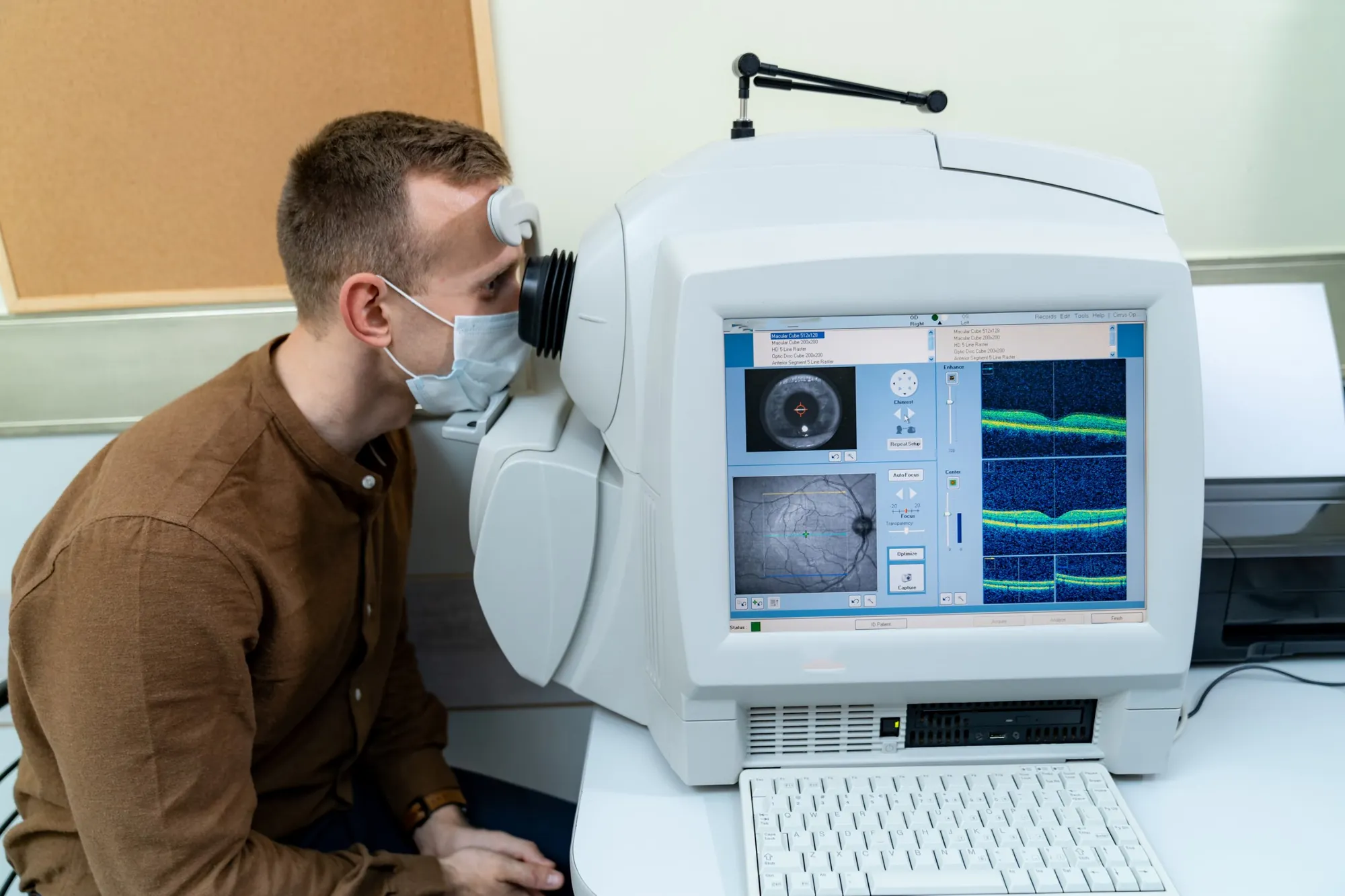
Diagnosis is made at a slit-lamp exam, with a look at the drainage angle (gonioscopy), a pressure check, a scan of the optic nerve (OCT) and a visual field test. Some clinics also scan the front of the eye to see the iris shape. Our guide to the glaucoma test explains each step. People with pigment dispersion but normal pressure and no damage need regular check-ups for life, and your eye doctor will decide how often. If your pressure starts to rise, checks are usually more frequent, often every 3 to 6 months.
Treatment lowers eye pressure to protect the optic nerve. It cannot stop pigment release entirely.
| Treatment | What to know |
|---|---|
| Eye drops | Usually first. In a 12-month trial of 36 people with pigmentary glaucoma, latanoprost lowered pressure by about 6 mmHg and timolol by about 5 mmHg |
| Pilocarpine | Flattens the iris and reduces pigment release, but blurs vision and causes brow ache, and is linked to retinal detachment, a particular concern in nearsighted eyes. Rarely used now |
| SLT laser | Lowers pressure about as well as in ordinary open-angle glaucoma, but pressure spikes after laser may be more common and the effect may fade within a few years |
| Laser iridotomy | Flattens the iris, but trials found no clear benefit in preventing glaucoma or field loss |
| Surgery | Trabeculectomy and drainage implants can control pressure well. In one 2026 study, a small implant (Preserflo) worked about as well as in ordinary open-angle glaucoma. Results with some MIGS stents have been mixed |
The same pressure-lowering drops used for other open-angle glaucoma are used here, most often a prostaglandin once a day. Because pigmentary glaucoma starts young, treatment may last decades, so side effects and the routine of daily use matter. Our glaucoma eye drops guide covers each type.
In selective laser trabeculoplasty (SLT), a laser treats the drainage tissue to help fluid flow out. Pigmented drainage tissue absorbs laser energy strongly, so doctors usually use lower energy. Even so, in a 2024 comparison of 65 matched eyes in each group, pressure spikes after SLT occurred only in eyes with pigmentary glaucoma (5 of 65 eyes, about 8%), though the laser worked about as well overall. In one small series treated over half the drain, success was 85% at one year, falling to 44% at three years. Your clinic will check pressure soon after laser. Read more in our SLT laser guide.
A tiny laser opening in the iris lets fluid pass, which flattens the backward-bowed iris and reduces rubbing. In theory this should help, but a Cochrane review of five trials found no clear benefit, with very low certainty. In the largest trial, of 116 people in the UK with pigment dispersion and high pressure, glaucoma developed in 15% of lasered eyes and 6% of untreated eyes over 3 years. The trial was too small to tell whether this difference was real or due to chance. It showed no benefit from the laser, and the need for drops was similar in both groups. One Italian trial found fewer pressure rises over 10 years in lasered eyes judged high risk on a dilating-drop test: 3 of 21 lasered eyes against 13 of 21 untreated eyes. No eye in that trial lost side vision, so it could not show that the laser protects sight. Some doctors may still consider it for younger people with marked iris bowing, but it is not a routine treatment.
When drops and laser are not enough, surgery can help. Trabeculectomy has controlled pressure well in long-term studies. In a 2026 study of 31 people with pigmentary glaucoma, a small drainage implant (the Preserflo MicroShunt) worked about as well as it did in matched people with ordinary open-angle glaucoma. Results with some minimally invasive glaucoma surgery (MIGS) stents have been mixed. Young nearsighted men may be more prone to very low pressure after surgery that makes a new drain (filtering surgery), so surgeons plan for this. Our guide to glaucoma surgery and laser covers the options.
With treatment, most people keep useful vision. In a Finnish study of 38 people with pigmentary glaucoma followed for about 10 years on average, 89% of eyes still had normal vision and 3% were blind, no one was blind in both eyes, and no one had to retire early because of it. Because it starts young, lifelong follow-up is the most important part of care.
Netra Eye Institute is an acupuncture and Oriental medicine practice. Nothing we offer lowers eye pressure, stops pigment release or replaces drops, laser or surgery. We found no study that has tested acupuncture or herbal medicine in pigment dispersion or pigmentary glaucoma, and a Cochrane review of acupuncture for glaucoma in general found the evidence too limited to draw reliable conclusions. Our guide to acupuncture and Chinese medicine for glaucoma explains what is and is not known.
Netra Restoration Therapy (NRT) does not treat glaucoma, lower eye pressure or restore any vision lost to glaucoma. For people whose glaucoma is medically controlled but who still struggle with field loss, low-vision optometrists and occupational therapists help with daily tasks. If you choose NRT, it is offered only alongside that care and your glaucoma treatment, never in place of them. Learn about Netra Restoration Therapy and low-vision rehabilitation.
No. Pigment dispersion syndrome means pigment is shedding from the iris. Pigmentary glaucoma means the shed pigment has helped raise eye pressure enough to damage the optic nerve. Most people with pigment dispersion syndrome do not develop glaucoma, but they need regular checks.
Usually not by itself, but it needs monitoring. In a US community study, about 15% of people developed glaucoma within 15 years. The risk was highest in people whose pressure was above normal at their first exam. Pigmentary glaucoma can cause permanent vision loss if untreated, so regular eye exams matter.
It often becomes less active with age, as the lens thickens and the iris moves away from the lens fibers, and some pigment can clear from the drain. It does not always settle, though, and any optic nerve damage is permanent. Keep up your eye exams even if your doctor says it has calmed down.
No. Like other glaucoma, it can be controlled but not cured, and vision already lost does not come back. Drops, laser and surgery lower eye pressure to stop or slow further damage, and many people keep good vision for many years with treatment.
Not unless your eye doctor advises it. Exercise can release pigment and briefly raise pressure in some people, but there is no evidence that it causes glaucoma. If you exercise hard and often, ask about a pressure check before and after your usual workout, and report blurred vision or halos after exercise.
It is not routinely recommended. The laser flattens the iris, but a Cochrane review and a 3-year UK trial found no clear benefit in preventing glaucoma or vision loss. Some doctors still consider it for selected younger people with marked iris bowing. Ask your eye doctor why they do or do not recommend it for you.
Sometimes. In an Iowa study, about 1 in 10 close relatives of people with the condition also had it, and some families pass it on in a dominant pattern. There is no routine genetic test, so relatives, especially nearsighted ones, should have regular eye exams.
Most people do not. In a Finnish study followed for about 10 years, 89% of treated eyes had normal vision and no one was blind in both eyes. The risk is highest when glaucoma is found late or not treated, so early diagnosis and regular follow-up are what protect your sight.
Pigment dispersion syndrome is found mostly in nearsighted young adults, and most people with it do not develop glaucoma. The risk is highest in people whose pressure is already raised when the condition is found. Regular exams, pressure-lowering treatment when needed, and caution with laser spikes keep most people seeing well. The condition often quiets with age, but follow-up should continue for life.
Medical Disclaimer: This article provides general education and is not medical advice, diagnosis or treatment. Pigment dispersion syndrome and glaucoma need assessment and follow-up by a licensed eye-care professional. Do not start, stop or change eye drops, laser plans or exercise habits for your eyes without your eye doctor. Severe eye pain, nausea or vomiting, or sudden vision loss needs urgent care. Integrative care at Netra Eye Institute does not lower eye pressure and must never delay or replace glaucoma treatment.