
Search conditions, treatments, research reviews and articles. Press Enter to see all results, Esc to close.
Search conditions, treatments, research reviews and articles. Press Enter to see all results, Esc to close.
Blog
August 9, 2026
Aerobic exercise, avoiding breath-holding during heavy lifting, not sleeping face-down and keeping caffeine moderate can each shift eye pressure a little. None lowers it enough to replace drops, laser or surgery, and none has been shown on its own to prevent field loss. Here is what the evidence supports, and by how much.
Living with glaucoma can generate two opposite reactions. Some people minimize a silent disease because they feel normal. Others try to control every meal, posture and moment of exercise after reading that an activity changes intraocular pressure for a few minutes. Neither approach reflects how glaucoma is managed.
The useful middle ground is structured and sustainable: use the treatment that has been shown to protect the optic nerve, keep monitoring appointments, support cardiovascular and metabolic health, and discuss the few activities that may need adjustment for your specific eyes. This guide separates outcome evidence from interesting but preliminary pressure observations.
Glaucoma is a group of optic neuropathies, not simply a high-pressure reading. Current treatment reduces intraocular pressure (IOP) because randomized trials show that doing so lowers the risk of developing or worsening damage. A normal office pressure does not guarantee stability, and a temporary decrease after a walk is not equivalent to long-term neuroprotection.
Lifestyle studies are difficult to interpret. Many measure IOP immediately after a single activity rather than visual-field progression over years. Participants, glaucoma types and treatments vary. People who exercise or eat well may also differ in income, access to care, smoking, diabetes or medication adherence. Association cannot establish that the habit caused a glaucoma outcome.
This does not make lifestyle irrelevant. Physical activity, nutritious food, smoking cessation, adequate sleep and treatment of systemic disease benefit the whole person. They may influence ocular perfusion, metabolic health, inflammation or pressure. The evidence boundary is that none should be sold as a substitute for drops, laser, surgery, OCT or visual-field monitoring.
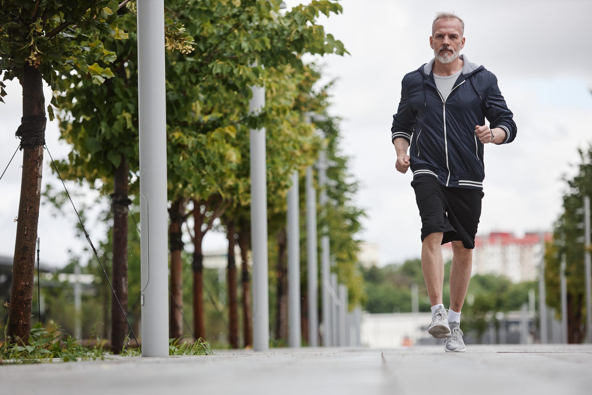
Most adults with stable glaucoma can follow public-health recommendations for regular aerobic activity and strength training, adjusted for their general health. Walking, cycling, swimming after surgical restrictions have ended, dancing and similar activities improve cardiovascular fitness, balance, mood and sleep. Maintaining strength is valuable for mobility and fall prevention when peripheral field is limited.
A 2024 systematic review found that aerobic and resistance sessions often produced an immediate IOP reduction in people with glaucoma. Longer-term results were inconsistent, and the authors emphasized small, heterogeneous studies. This supports exercise as a healthy behavior, not a prescription to replace an ophthalmic treatment.
Exercise also affects blood pressure, hydration and breathing. A short IOP measurement after exertion captures only one part of optic-nerve physiology. The outcome that matters is whether structure and field remain stable over time.
Moderate aerobic activity commonly causes a temporary IOP decrease. People who are sedentary can start gradually, such as ten-minute walks, and build toward a clinician-approved plan. Choose a setting that accommodates field loss: even pavement, good lighting, handrails, a companion or a stationary machine may reduce collision and fall risk.
Advanced glaucoma does not automatically prohibit exercise. It may change the safest format. Someone who misses lower steps may benefit from orientation-and-mobility assessment, contrasting stair edges and an exercise area without moving hazards.
Resistance training is important for bone, muscle and metabolic health. IOP can rise transiently during heavy effort, especially with a Valsalva maneuver—straining while holding the breath. Higher loads, longer effort and some body positions may create larger changes.
For many people, practical modifications are to use a manageable load, breathe continuously, avoid prolonged maximal straining and obtain instruction on form. This is not evidence that every glaucoma patient must lift only tiny weights. A glaucoma specialist may give more conservative advice for an eye with severe central threat, very unstable pressure or recent filtering surgery.
Head-below-heart positions can raise IOP while the posture is held. Examples include prolonged full inversions and some inverted yoga poses. The pressure usually changes again after returning upright, and the long-term effect on progression is not well established.
People with glaucoma can ask an instructor for upright modifications rather than abandoning yoga’s flexibility, balance and stress benefits. A brief forward bend is not identical to a sustained headstand. Disease severity, surgical history and tolerance matter.
Tight small-cup goggles can raise periocular and sometimes measured IOP while worn. Comfortable, properly fitted goggles are sensible. Swimming may be restricted temporarily after laser or surgery because of infection and wound concerns; the operating surgeon’s instructions control.
Routine commercial air travel is usually possible with glaucoma, but an intraocular gas bubble placed for retinal surgery is a different emergency risk and prohibits altitude exposure until cleared. Glaucoma implants themselves are not gas bubbles. High-altitude travel, hypoxia and pressure questions should be individualized for advanced disease and cardiopulmonary conditions.
No glaucoma diet has been shown to restore retinal ganglion cells. Observational studies have explored leafy vegetables, fruit, tea, coffee, fats and dietary patterns, but results are not consistent enough to prescribe a food as glaucoma treatment.
A Mediterranean-style or similarly balanced pattern is reasonable for cardiovascular and metabolic health: vegetables, fruit, legumes, whole grains, nuts, fish or other appropriate proteins, and limited highly processed food. This can support diabetes, blood pressure and weight management. Its value does not depend on claiming that one nutrient reaches the optic nerve and reverses damage.
People with kidney disease, anticoagulation, diabetes, allergies or other dietary restrictions need individualized guidance. “Natural” foods and products can interact with medicines or contain unsafe doses.
Caffeine can produce a modest, temporary IOP increase in some people. The response varies with dose and habitual use. Evidence does not support a universal ban on coffee for every person with glaucoma.
Avoid using a large caffeine dose immediately before a pressure check if the clinician wants a representative baseline, and discuss unusually high intake or sensitivity. Decaffeinated options and smaller servings are available. The goal is proportionality, not fear of a morning cup.
Rapidly drinking a large volume of water can transiently raise IOP and is sometimes used in research as a provocative test. Normal hydration spread through the day is different. Dehydration can cause dizziness, kidney problems and low blood pressure and is not a glaucoma therapy.
Fluid limits prescribed for heart, kidney or endocrine disease take priority. Do not manipulate hydration to change an eye-pressure number.
Alcohol may transiently alter pressure, but it is not treatment. Regular or heavy use carries major health and safety risks and can worsen sleep and medication consistency. Follow general medical guidance rather than drinking for an ocular effect.
Smoking harms cardiovascular health and is associated with multiple eye diseases. The precise relationship with glaucoma progression is less consistent than for some other conditions, but cessation remains a high-value health action. Evidence uncertainty is not a reason to continue tobacco.
Antioxidants, ginkgo, citicoline, nicotinamide, saffron and many proprietary combinations are marketed for optic-nerve protection. Laboratory mechanisms, a biomarker change or a small short trial cannot establish prevention of disability. Products vary in dose and purity, and supplements do not pass through the same approval pathway as glaucoma drugs.
High-dose nicotinamide has generated research interest, but doses used in trials can affect the liver, gastrointestinal system, glucose metabolism and other organs. It should not be self-prescribed as a benign vitamin. Ginkgo can increase bleeding risk and interact with anticoagulants or surgery. Supplements may also duplicate ingredients across bottles.
AREDS2 is formulated for selected stages of age-related macular degeneration, not glaucoma. Taking it because the label says “eye health” does not treat the optic nerve.
Before starting a product, ask what human outcome it improved, in which glaucoma type, for how long, at what dose and with what adverse-event monitoring. Share every supplement with the ophthalmologist, primary clinician and pharmacist.
Cannabinoids can lower IOP for a few hours in some people. Glaucoma requires control around the clock for years. Maintaining an effect would demand frequent exposure, while impairment, dependence, psychiatric effects, lung exposure with smoking and changes in blood pressure may compromise safety or optic-nerve perfusion.
Cannabis also impairs driving and can interact with other medicines. No major ophthalmic treatment strategy recommends it in place of longer-acting, tested pressure therapies. A short pharmacologic effect is not the same as a practical treatment.
IOP and blood pressure change across the day and with body position. Pressure is often higher when lying down than sitting. Some people also experience substantial nocturnal blood-pressure lowering. The relationship among nighttime IOP, perfusion and glaucoma progression is complex; a single office measurement cannot reconstruct it.
Good sleep supports cognition, medication routines, mood and systemic health. Persistent insomnia, loud snoring, witnessed pauses, morning headaches or daytime sleepiness merit medical assessment. Obstructive sleep apnea has been associated with glaucoma in some studies, but association and treatment effects remain complex.
Continuous positive airway pressure protects people with clinically important sleep apnea from established cardiopulmonary and daytime consequences. Some research suggests CPAP can alter nighttime IOP. That is a reason for coordination and monitoring, not a reason to abandon therapy independently.
Tell the glaucoma specialist about sleep apnea and the sleep clinician about glaucoma. They can review disease severity, CPAP effectiveness, systemic pressure and monitoring. Untreated hypoxia and cardiovascular risk can be dangerous.
The dependent eye may have higher pressure during side sleeping, and head elevation can affect nocturnal IOP in experimental settings. Long-term evidence that prescribing one sleep position prevents visual-field loss is limited.
Someone with asymmetric advanced glaucoma can ask whether position is relevant, but sleep quality, reflux, spine disease, fall risk and apnea also matter. Stacking unstable pillows or losing sleep in pursuit of a small pressure change may cause more harm.
Blood pressure that falls very low at night has been investigated as a progression factor, particularly in normal-tension glaucoma. However, hypertension treatment prevents stroke, heart attack and kidney disease. Moving or withholding medicine without supervision can be dangerous.
If progression continues at low IOP, the ophthalmologist may communicate with the prescribing clinician and consider ambulatory blood-pressure monitoring. Decisions should use actual data and overall cardiovascular risk.
Glaucoma drops are unusually difficult to value from day to day: they generally do not sharpen sight or create a sensation that confirms they worked. Their benefit is a lower probability of future loss. This makes a reliable routine more important than waiting to feel a difference.
Know the name, cap color, dose and eye for every bottle. Keep an updated list on the phone and in a wallet, because appearance can change with generic manufacturers. At each visit, reconcile what the chart says with what is actually being used. “One drop at night” is incomplete if the household contains several bottles.
Wash and dry hands. Check the label. Tilt the head back or lie down, pull the lower lid gently to make a pocket, look up and place one drop without touching the tip to the eye, lashes, fingers or skin. Close the eye gently rather than squeezing repeatedly. If instructed, press near the inner corner for a minute or two to reduce drainage into the nose.
One successful drop is enough; the conjunctival sac cannot hold a stream. If uncertain whether any drop entered, follow the prescriber’s advice—one extra drop is usually less concerning than routinely missing treatment, but repeated flooding increases waste and exposure. Separate different medicines by at least several minutes so the second does not wash out the first.
Bottle aids, a mirror, lying flat, refrigeration when labeling permits, caregiver assistance or single-use preservative-free units may help. Never share bottles. Replace a bottle whose tip is contaminated, and follow storage and expiration instructions.
Link dosing to a stable cue such as brushing teeth, breakfast or a phone alarm. Keep medication where it will be seen but protected from children, heat and contamination. Travel with it in carry-on luggage and preserve required temperature conditions.
A weekly checklist can identify patterns without creating shame. Pharmacy synchronization and automatic refills reduce gaps. If work hours or caregiving makes a dose unrealistic, say so; another class, fixed combination or SLT may produce a more durable plan.
Do not double doses automatically after a missed one. Product instructions and timing differ. Ask the care team or pharmacist for a written missed-dose rule.
Redness, burning, dryness, lash growth, lid or iris changes, allergy, fatigue, slow pulse, breathing difficulty, dry mouth and altered taste can occur depending on the class. New shortness of breath, faintness or a severe reaction requires prompt medical advice.
Surface symptoms may improve with a preservative-free formulation, fixed combination, lubricants, treatment of lid disease, SLT or a different agent. Artificial tears should be spaced from prescription drops. Steroid eye drops are not casual redness remedies because they can raise pressure and cause other complications.
If cost is the problem, contact the practice before rationing. Formularies, generics, prior authorization, assistance programs and procedures can change the plan. The clinician cannot solve a barrier that remains hidden.
Pressure checks alone cannot prove that glaucoma is stable. Follow-up may include gonioscopy, optic-nerve examination, photography, OCT and visual fields. The interval depends on stage, target, rate and treatment change.
Bring glasses and an updated medication list. Use drops normally unless specifically told otherwise; withholding them to “show the real pressure” can create an unsafe and uninterpretable visit. Mention steroid exposure from any route, emergency visits, falls, driving concerns and new neurologic symptoms.
Visual fields are effort-dependent, but perfection is not required. Ask for positioning or a pause if uncomfortable, keep looking at the central target and press only when a stimulus is seen. Repeat testing is common because clinicians distinguish learning, fatigue and artifact from true change.
Home tonometry can reveal patterns in selected patients, yet technique, device agreement and data overload are limitations. It should answer a defined clinical question. Consumer wearables and phone apps do not diagnose glaucoma or replace calibrated testing.
Glaucoma belongs on every medication history. Steroids delivered as eye drops, pills, inhalers, injections, nasal sprays or skin products can raise IOP in susceptible people, with response varying by potency, route, duration and individual biology. Steroids may still be essential for asthma, autoimmune disease, transplant care or postoperative inflammation. The safe approach is to tell both clinicians and arrange pressure monitoring—not to discontinue an important medicine independently.
Some cold, allergy, bladder, motion-sickness and psychiatric medicines carry warnings about glaucoma because anticholinergic or adrenergic effects can precipitate angle closure in anatomically narrow angles. This warning does not mean every person with open-angle glaucoma must avoid every product. Ask what type of glaucoma and angle anatomy you have, then let the pharmacist and ophthalmologist evaluate the specific ingredient.
Systemic beta blockers, carbonic-anhydrase inhibitors and other medicines can overlap with glaucoma therapy. Bring a complete list to dental procedures and urgent care as well as eye visits. Complementary products count as medicines when considering bleeding, blood pressure, sedation and surgery.
Protective eyewear remains valuable. Eye trauma can cause angle recession and secondary glaucoma months or years later. Use sport- or task-appropriate impact protection for racquet sports, workshop tools, yard work and occupations with projectiles or chemicals. Ordinary sunglasses are not safety glasses.
Contact lenses can complicate drop timing, surface comfort and bleb safety. Many drops should be instilled after lenses are removed, with a labeling-specific wait before reinsertion. A filtering bleb, recent operation, redness or epithelial disease may change whether contacts are appropriate. Obtain direct instructions instead of applying a generic timetable.
Screens do not cause glaucoma. They can produce dryness, fatigue and postural discomfort, particularly when blink rate falls. Breaks, appropriate correction, larger text and lubrication can improve comfort, but a blue-light filter does not lower IOP. Screen adaptations become rehabilitative when field or contrast loss makes navigation difficult.
Travel planning should include extra medicine in original labeled packaging, a copy of prescriptions and the treating practice’s contact information. Time-zone changes are easier with a written dosing plan. Do not pack the only supply in checked luggage, and do not expose products to temperatures outside their label. Identify urgent eye care in advance for remote travel after recent surgery.
These precautions are most useful when they simplify care and prevent interruptions, not when they turn ordinary activity into a source of constant alarm.
Glaucoma may affect contrast, adaptation and areas of peripheral vision before central acuity falls. A person can read an eye chart well yet clip doorframes, miss a curb or struggle in dim environments. These experiences deserve functional evaluation, not dismissal because central acuity is “20/20.”
Improve lighting while controlling glare. Use contrast on stair edges, cutting boards, switches and medication labels. Keep walkways predictable, remove loose rugs and place frequently used objects consistently. Scan deliberately at intersections, in stores and before stepping down.
Driving eligibility is determined by local law and functional testing, not diagnosis alone. Glare, night conditions and field defects can affect safety. Discuss near misses honestly and consider a driving rehabilitation evaluation when appropriate. Stopping driving can be emotionally and practically difficult; transportation planning should begin before a crisis.
At work, screen magnification, larger pointers, contrast settings, task lighting and workspace organization may help. Disability and rehabilitation services can address job-specific needs. These accommodations improve access; they do not indicate disease progression or treatment failure.
A chronic, initially silent disease asks people to accept treatment today for a benefit they may never directly feel. Fear of blindness, frustration with fields, surgical anxiety and guilt about missed doses are common. Advanced loss can reduce independence and increase depression or social isolation.
Accurate information can reduce catastrophic thinking: treated glaucoma does not inevitably cause blindness, but established damage deserves respect. Ask the clinician to explain stage, rate, target and what has remained stable. That is more meaningful than comparing one IOP number with another patient’s.
Support groups, counseling and low-vision services are appropriate before total or severe loss. Care partners can help with drops and transportation while preserving autonomy. If anxiety or depression affects sleep, function or adherence, involve primary or mental-health care.
Procedure-specific instructions override general lifestyle advice. After SLT, cataract/MIGS, trabeculectomy or tube surgery, restrictions on water exposure, rubbing, lifting, bending, makeup, contact lenses and exercise differ. Filtering surgery often requires frequent early visits and careful wound management.
Do not decide that a clear eye or better vision means postoperative drops can stop. Steroids and antibiotics have schedules; glaucoma drops may differ between operated and fellow eyes. Written calendars reduce errors.
Worsening pain, redness, discharge, sudden blur, flashes, a curtain, nausea or vision loss requires urgent contact. A filtering bleb carries a lifelong infection risk, so late redness and pain still matter. Low pressure can be dangerous as well as high pressure.
A sustainable plan might include:
The plan should be short enough to live with. If it consumes the day, ask which actions actually change outcomes.
At Netra Eye Institute, NRT is supportive rehabilitation. It cannot lower IOP, replace prescribed drops, substitute for SLT or surgery, reopen a closed angle, reverse OCT thinning or regenerate retinal ganglion cells. A person whose pressure or field is unstable needs ophthalmic treatment, not a rehabilitation delay.
After the glaucoma specialist confirms medical stability, established loss may create problems with visual search, scanning, reading fluency, contrast, crowded environments or mobility. A functional evaluation can define a measurable task goal and identify whether structured practice, environmental modification, low-vision devices or referral to orientation-and-mobility services is appropriate.
Success should be described as better task performance, strategy use or confidence—not restoration of the missing visual field. Pressure, OCT and standardized perimetry remain the measures of disease control.
Learn about Netra Restoration Therapy, Netra Eye Institute’s approach, glaucoma testing and living with low vision.
A walk may temporarily lower IOP and benefits general health. It has not been shown to replace prescribed pressure treatment or monitoring.
Usually not. Avoid breath-holding and prolonged maximal strain, and ask for individualized restrictions if disease is advanced or surgery is recent.
No universal ban is supported. Large caffeine doses can temporarily raise IOP in some people, so discuss heavy intake or unusual sensitivity.
No vitamin has been proven to cure glaucoma or restore the optic nerve. High-dose supplements can cause harm and should be reviewed clinically.
Not routinely. Position can change IOP, but evidence that a prescribed posture prevents progression is limited. Sleep quality and other medical needs matter.
No. Untreated sleep apnea has serious risks. Coordinate the sleep and glaucoma teams so both conditions are monitored.
They are preventive: lowering pressure reduces the chance of future damage. They generally cannot restore vision already lost.
No. Stability is achieved and assessed through medical pressure control, OCT, fields and examination. NRT may support selected functional goals after stability.
Everyday glaucoma care is less about finding a miracle habit than making proven care workable. Exercise, balanced food, sleep care, smoking cessation and systemic health support the person; medication technique, affordability and monitoring directly support the glaucoma plan. None replaces pressure lowering.
Use lifestyle evidence in proportion. A temporary IOP change is not a cure, and uncertain research should not provoke unsafe changes to blood-pressure medicine, CPAP, hydration or nutrition. Once medical care is secure, rehabilitation—including NRT in appropriately selected cases—can focus on living better with function that remains.
Sustainable routines matter more than occasional perfect days. Link drops and appointments to reliable cues, then revise the system when work, travel, dexterity or memory changes.
Medical Disclaimer: This article provides general education and is not medical advice, diagnosis or treatment. Do not change glaucoma drops, blood-pressure medicines, CPAP, supplements, hydration, exercise restrictions or postoperative care without the relevant licensed clinicians. Sudden eye pain, redness, blurred vision, halos, nausea, vomiting or vision loss requires emergency evaluation. NRT must never delay or replace pressure-lowering care.



