
Search conditions, treatments, research reviews and articles. Press Enter to see all results, Esc to close.
Search conditions, treatments, research reviews and articles. Press Enter to see all results, Esc to close.
Blog
August 9, 2026
Cupping is the hollowed-out look of an optic nerve that has lost nerve fiber tissue, measured as the cup-to-disc ratio. A large cup alone does not mean glaucoma; some nerves are simply born big. What counts is the pattern, the neuroretinal rim, and whether it changes over time. This page explains what your scan shows.
Glaucoma testing can feel contradictory. A pressure is “normal,” yet an OCT sector is red. A visual field is worse, but the optic nerve photograph looks unchanged. One clinician calls a person a suspect; another recommends treatment.
These differences do not necessarily mean the tests are poor or the clinicians disagree about basic facts. Glaucoma is a longitudinal diagnosis: pressure is exposure, OCT and photographs measure structure, fields measure function, and angle examination identifies mechanism. Each samples a different part of the disease.
This guide explains how those pieces fit without duplicating Netra Eye Institute’s glaucoma condition pages. It emphasizes what the tests reveal, how they fail and how a baseline becomes a progression decision.
Testing should answer a specific question:
The same test can have a different role at baseline and follow-up. A screening OCT may flag risk; serial OCT estimates rate. A field confirms function and later determines whether loss is accelerating. More tests are not automatically better unless they change the answer or increase confidence.
Open-angle glaucoma is usually asymptomatic early. The clinician therefore asks about family subtype and blindness, steroid exposure, trauma, uveitis, surgery, myopia, sleep apnea and systemic medications rather than waiting for symptoms.
Functional history still matters: collisions, missed steps, driving concerns, one-sided search difficulty, reading interruptions and difference between eyes may reveal impact not summarized by acuity. Sudden pain, redness, nausea or rapid field loss suggests an emergency or another condition.
Bring prior records, because change is central. An isolated cup-to-disc ratio or pressure from years ago is less useful than the images and fields themselves.
Visual acuity tests central high-contrast detail. It can remain 20/20 in advanced peripheral glaucoma. Reduced acuity prompts consideration of central field involvement, cataract, corneal disease, retina or neurologic causes.
Refraction determines how much blur is optical. Pupils are checked for a relative afferent defect that may appear with asymmetric advanced optic-nerve damage but is not specific to glaucoma.
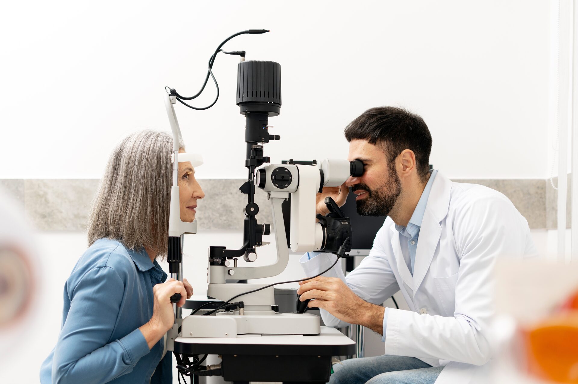
The slit lamp evaluates cornea, anterior chamber, iris, lens and signs of pseudoexfoliation, pigment dispersion, inflammation, trauma or prior surgery. Corneal edema can indicate acute pressure elevation. Lens and ocular-surface findings explain visual complaints and test quality.
Goldmann applanation tonometry estimates pressure from the force needed to flatten a defined corneal area and is the clinical reference standard. Other methods include rebound, pneumatonometry, dynamic-contour devices and noncontact air-puff screening.
Measurements differ because devices use different physics and corneal assumptions. Time of day, breath holding, eyelid squeezing, tight collars, recent exercise, body position and technique can alter results. The chart should record method when comparisons matter.
It can identify a very high value requiring action, document response to treatment and contribute to risk. It cannot show whether retinal ganglion cells are already damaged, identify the angle mechanism or define the day’s peak.
Target is an estimated range expected to slow damage based on baseline IOP, stage, rate, age, central field and other risk. It is not a magic universal number. The target can be revised when testing shows progression or stability.
IOP varies across 24 hours. Office curves or prescription rebound self-tonometry may reveal timing and peaks for selected patients. Home data can be noisy from technique and may increase anxiety. It is useful only when the clinician has a question and a plan for interpreting results.
Do not press on the eye, use another person’s device without training or alter drops from a home reading.
Ultrasound or optical pachymetry measures central corneal thickness. A thinner cornea can produce a lower Goldmann reading than a thicker cornea under some conditions and was a conversion-risk marker in ocular hypertension research.
Thickness is not the only biomechanical property. Rigidity, hysteresis, curvature, surgery and disease affect measurement. Fixed online correction tables falsely imply that adding or subtracting a number yields the true pressure.
Corneal refractive surgery can lower measured applanation IOP without lowering actual glaucoma risk. Prior LASIK or PRK and preoperative records should be disclosed. Alternative tonometry and greater reliance on nerve and field trends may be needed.
The cornea–air interface prevents a direct view of the anterior chamber angle. A gonioscopy lens neutralizes that interface and uses mirrors to show the trabecular meshwork, scleral spur and other structures.
Gonioscopy can identify:
Indentation gonioscopy helps distinguish appositional closure from permanent adhesions. Lighting and pressure on the lens affect the view, so technique matters.
Anterior-segment OCT or ultrasound biomicroscopy can quantify angle and lens–iris configuration, especially when anatomy is complex. They complement, not universally replace, dynamic gonioscopy.
The clinician assesses disc size, neuroretinal rim, retinal nerve fiber layer, peripapillary atrophy, asymmetry and disc hemorrhage. Glaucomatous loss often produces focal rim notching or thinning that matches nerve-fiber anatomy.
Large discs can have large physiologic cups; small discs can look crowded despite damage. High myopia creates tilt, torsion and peripapillary changes. Pallor greater than cupping, severe acuity or color loss, unusual laterality or a vertically respecting field suggests a nonglaucomatous optic neuropathy.
The cup-to-disc ratio compresses complex anatomy into one number and should not be used alone. Dilation also examines retina for macular, vascular or peripheral causes of field loss.
Stereoscopic or high-quality color photography provides a durable baseline for rim, vessels, hemorrhage and peripapillary tissue. Photographs are not limited by a normative database and can remain interpretable when OCT reaches a floor.
Progression may appear as focal rim loss, vessel position change or new hemorrhage. Image alignment, focus and disc size matter. Side-by-side comparison across years is more useful than relying on memory.
Photography samples visible structure but may miss subtle diffuse loss. It works alongside OCT and fields.
Spectral-domain OCT measures peripapillary retinal nerve fiber layer (RNFL) thickness around the disc. Glaucomatous loss often follows superior and inferior arcuate bundles, producing corresponding field defects.
The report compares measurements with an age-matched reference and displays green, yellow or red classifications. These colors are statistical, not diagnostic. “Red disease” occurs when healthy anatomy falls outside the database; “green disease” occurs when a real focal defect remains within broad normal limits.
Clinicians inspect scan centration, signal, segmentation and the thickness curve. Media opacity, blinking, vitreous traction, epiretinal tissue, blood vessels and myopia can alter measurement.
In advanced glaucoma, RNFL thickness approaches residual non-neural tissue and changes little despite further functional loss. Macular measures and visual fields may remain informative. A stable floor-level OCT does not prove stability.
About half of retinal ganglion cells are associated with the macular region. OCT analysis of ganglion cell–inner plexiform layer or ganglion cell complex can reveal central or paracentral loss and complement peripapillary RNFL.
Macular disease, epiretinal membrane, edema, high myopia and segmentation errors can distort results. A central defect should be correlated with a 10-2 or denser field and a macular structural scan.
Asymmetry maps and probability plots help identify patterns, but software names and boundaries differ among devices.
Bruch’s membrane opening–minimum rim width and three-dimensional rim measurements attempt to quantify the shortest tissue path at the disc margin. Swept-source systems provide deeper or wider imaging. OCT angiography measures vessel-density signals.
These tools can add information, particularly in challenging discs, but none independently proves glaucoma or progression. Vessel density is influenced by signal, segmentation, systemic physiology and existing tissue loss. Whether a vascular change is cause or consequence can be unclear.
Artificial-intelligence algorithms can classify images with high performance in research datasets, while progression prediction and external generalization are less robust. A probability score requires clinician review.
Visual-field testing measures light-detection sensitivity at sampled locations while the patient fixates centrally. The bowl or screen varies stimulus brightness, and responses are compared with age-based expectations.
Common patterns include 24-2, 30-2 and 10-2. A 24-2 samples much of the central field used for routine glaucoma care; 10-2 densely samples the central ten degrees. The 24-2C adds selected central locations while retaining a broader grid.
Perimetry is a psychophysical test: it measures a person’s responses, not a photograph. This does not make it subjective or optional. It is the primary clinical measure of how glaucoma affects visual function.
Wear the correction provided, sit comfortably, keep looking at the fixation target and press only when a light is seen. Blinking is allowed; ask to pause if the eye is dry, posture is painful or instruction is unclear. Do not chase lights with the eyes or try to predict timing.
The first test is often less reliable because the task is unfamiliar. Improvement on repeat may represent learning rather than recovery.
Fixation losses, false positives and false negatives help judge quality but are not simple pass–fail rules. False-positive responses can create artificially good sensitivity and unusual patterns. False negatives increase with advanced loss, fatigue and fluctuation.
Gaze tracking, technician observation and the pattern of responses add context. A test with one flagged index can still contain useful information; a visually smooth test can still be wrong.
Total deviation compares each point with age-matched normal sensitivity. Pattern deviation adjusts for generalized depression such as cataract to highlight localized defects, though in advanced loss this adjustment can mislead. Probability symbols indicate statistical rarity, not certainty of glaucoma.
Mean deviation summarizes overall difference; pattern standard deviation reflects localized irregularity; the visual field index is another global summary. Two eyes with the same global index can have very different defect locations and functional impact.
Event analysis asks whether change exceeds expected test–retest variability compared with baseline. Trend analysis estimates a slope over time. Both require enough reliable tests.
A statistically significant slope is not automatically clinically threatening. Rate must be interpreted against age, baseline stage, central involvement and life expectancy. Conversely, a modest global slope can hide a rapidly changing central cluster.
Testing more frequently early after diagnosis helps estimate rate, but burden and learning matter. The clinician chooses intervals based on uncertainty and risk rather than a universal annual schedule.
Structure and function are related but not synchronized. OCT may detect early thinning before a field defect becomes repeatable. Later, OCT can reach a floor while fields continue to worsen. Test noise and different sampling locations further reduce same-visit agreement.
A 2024 longitudinal study found that structural tests and fields identified progression at the same visit in only a minority of progressing eyes. This supports multimodal follow-up rather than choosing a winner.
When tests disagree, clinicians ask:
Treatment may be adjusted when one convincing modality shows progression, especially in a high-risk eye. Agreement is reassuring but not required for real change.
A baseline is not one visit. Pressure varies, fields have learning effects and image quality differs. Early repeat testing establishes reproducibility and the patient’s range.
Useful baseline material includes gonioscopy, pachymetry, disc photographs, at least two reliable fields when feasible, OCT RNFL and macular ganglion cells, medication timing and prior untreated pressure. Each eye is staged separately.
After treatment begins, baseline pressure and structure still matter. A lower number has meaning only relative to the untreated exposure and subsequent stability.
Cataract reduces the amount and quality of light reaching the retina. On fields it can produce generalized depression and worsen global indices without a new localized glaucomatous defect. After cataract surgery, sensitivity may improve, creating an apparent break in the trend. Clinicians annotate lens status and interpret pre- and postoperative baselines carefully.
Cataract also lowers OCT signal and can introduce segmentation error. A better scan after surgery may measure thicker RNFL simply because the boundary is clearer, not because nerve fibers returned.
Dry eye causes fluctuating acuity, discomfort and blinking during perimetry. It can reduce OCT signal and make long testing sessions difficult. Lubrication approved by the clinician, rest breaks and scheduling can improve quality. Preserved glaucoma drops may contribute to ocular-surface disease; treating it improves adherence and test reliability without treating optic neuropathy.
Corneal scars, edema and contact lenses affect tonometry and optical imaging. The examination should document the limitation rather than force a precise number from poor conditions.
Axial elongation stretches peripapillary tissue and changes the normal RNFL bundle trajectory. Tilted discs, peripapillary atrophy and scan-circle placement can generate red sectors in a healthy eye. Macular schisis or myopic degeneration may alter ganglion-cell maps and fields.
Clinicians inspect the actual B-scans, compare both eyes cautiously and emphasize serial change. Myopia-specific normative databases and wide-field maps can help but are not infallible. Disc photographs remain valuable.
A field defect from myopic maculopathy or tilted-disc syndrome may not follow a typical glaucomatous pattern. Glaucoma can still coexist, so “it is just myopia” and “every red sector is glaucoma” are equally unsafe shortcuts.
In advanced disease, standard OCT RNFL approaches a floor and 24-2 fields may contain many points with high variability. Denser central fields, larger stimulus sizes in selected protocols, macular OCT, photographs and careful clinical examination can extend the useful range.
Global indices can hide a small remaining central island. Progression threatening fixation deserves attention even when mean deviation changes little. Testing should prioritize the remaining field relevant to function.
Fatigue and false-negative responses increase with severe loss because near-threshold lights are genuinely difficult. Reliability must be judged in context rather than excluding every hard test.
At the opposite extreme, a glaucoma suspect may need repeated baseline fields before a subtle defect is trusted. Test strategy changes with stage.
Optic neuritis, ischemic optic neuropathy, compressive lesions, hereditary neuropathy, retinal vascular disease and neurologic injury can produce cupping or field loss. Red flags include disproportionate optic-disc pallor, reduced color and acuity early, pain with eye movement, rapid change, a vertical-meridian defect or poor structure–function correspondence.
OCT can show ganglion-cell loss from neurologic disease, sometimes in a pattern corresponding to the visual pathway. A machine glaucoma classification does not identify the cause. Neuro-ophthalmic examination and imaging may be required.
Medication toxicity and retinal degeneration can also produce ring or central defects. Dilation, macular OCT, autofluorescence or electroretinography may redirect diagnosis.
The label should follow the anatomy. Treating pressure is appropriate when glaucoma coexists, but it cannot solve a compressive or retinal problem.
A field slope may be expressed in decibels per year, while OCT uses micrometers per year. Both are abstractions with stage-dependent measurement ranges. Rate becomes clinically meaningful when projected against remaining function and expected lifetime.
An older patient losing a small amount slowly and a younger patient losing rapidly can have the same current field but different urgency. Central location raises stakes. Confidence intervals matter: a slope based on three noisy visits is less certain than one based on repeated high-quality tests.
Clinicians may increase test frequency when a rate appears fast, then lower target pressure if confirmed. A single event alert should be repeated unless the change is clinically obvious or risk makes waiting unsafe.
Treatment itself changes the future trajectory. Extrapolating a pre-treatment slope indefinitely after surgery or major pressure reduction is inappropriate; a new trend must be established.
Ask for a summary that includes subtype, stage in each eye, angle status, highest known and target pressure, current medicines or procedures and last reliable field/OCT dates. A traffic-light color printout without interpretation is not enough.
When changing practices, transfer disc photographs, original field reports and OCT progression pages if possible. Raw OCT files may not import across manufacturers, but images and numerical tables preserve context.
Record allergies and intolerances separately from medications that simply did not achieve target. Note refractive and cataract surgery dates because they alter measurement comparability.
A concise record helps during travel, hospitalization and emergencies and prevents a new normal pressure from being mistaken for untreated baseline.
Good longitudinal care uses the same device and scan registration when possible. Changing OCT manufacturers can interrupt trend analysis because segmentation and reference definitions differ. Bring printed reports or raw exports when changing practices.
Clinicians compare global and sectoral slopes, photographs, field clusters and central points. They also review cataract surgery, macular change and test reliability. Progression should be documented with a rate and location, not only the statement “worse.”
An eye can remain stable because treatment works; stability does not prove the original diagnosis was wrong. Conversely, a slowly changing suspect may need reclassification.
Prescription rebound tonometers can measure IOP outside office hours. Tablet, computer and virtual-reality perimetry may allow more frequent field sampling. Portable cameras and tonometers can extend screening in underserved settings.
Home testing faces calibration, fixation, lighting, refractive correction, adherence and digital-access challenges. Frequent low-quality tests create false alarms or noise. Devices should transfer results to a clinical team with defined review and escalation.
A 2026 review concluded that tabletop perimetry is better positioned to complement standard automated perimetry than replace it. Smartphone screen size limits comprehensive field coverage. Consumer apps should not drive medication changes.
A screening program identifies people who need comprehensive evaluation. An air puff, nonmydriatic photo or portable field can improve access but will miss some disease and flag some healthy eyes.
NEI specifically notes that tonometry alone is insufficient because normal pressure occurs in glaucoma and high pressure occurs without it. A failed screening is not a diagnosis; a passed screening is not lifelong clearance.
Referral pathways determine whether screening prevents blindness. Programs need timely confirmatory care, language access and follow-up for ungradable or suspicious results.
Use prescribed drops normally unless instructed otherwise and note the last dose. Bring medication bottles, prior records and current glasses. Tell staff about steroid use, trauma, refractive surgery and difficulty with prior fields.
Sleep and eat normally. Caffeine or exercise should not be manipulated unless the clinician is testing a specific question. Ask for ergonomic adjustment or breaks; pain and fatigue reduce quality.
Afterward, ask:
Request a plain-language explanation of the image rather than relying on its color.
Pressure is one risk measure. Normal-tension glaucoma and fluctuating pressure make the statement unsafe.
Reference colors can be false-positive in myopia or unusual anatomy. The raw image, disc, field and trend determine meaning.
Technicians and clinicians assess reliability. Repeat testing may be necessary, but a difficult test can still show a consistent pattern.
Advanced OCT can reach a floor, and function may change first. Fields and photographs remain necessary.
It may improve frequency but still requires examination, calibrated devices and clinician review. An alert may create an extra visit.
NRT at Netra Eye Institute cannot measure IOP, inspect the drainage angle, diagnose optic neuropathy or determine progression. It does not replace OCT, fields, drops, laser or surgery.
After glaucoma is medically treated and stable, task difficulty may not be captured by global field indices. A functional assessment can examine search strategy, scanning, contrast, reading, eye–head coordination, divided attention and endurance.
Training can target use of the remaining field during selected tasks, but an improved search score does not mean retinal ganglion cells regenerated or the Humphrey field expanded. Medical and functional outcomes must remain separate.
Learn about Netra Restoration Therapy, Netra Eye Institute’s approach and low-vision rehabilitation.
No. It measures pressure but not the nerve, angle or field. Comprehensive evaluation is needed when risk or findings are concerning.
Angle anatomy determines mechanism and acute-closure risk. Narrow or scarred angles can exist without a high reading at that moment.
Frequency depends on stage, rate uncertainty and risk. Several early tests may establish a slope; stable low-risk disease may need less frequent testing.
Sometimes. Structural loss can precede repeatable perimetric damage. The reverse can occur in advanced disease or when imaging is limited.
Most people tolerate dilation. Eyes with occludable angles require individualized assessment. Acute pain, blur, halos or nausea after dilation needs urgent evaluation.
AI can support detection, but performance varies outside training data and it cannot replace angle, pressure, history or longitudinal judgment.
No. NRT may improve selected task strategies after stabilization; it does not thicken the RNFL or restore the optic nerve.
Glaucoma testing is a coordinated comparison of exposure, anatomy, structure and function. Pressure and cornea establish context; gonioscopy identifies mechanism; examination and photography show the nerve; OCT quantifies layers; perimetry measures vision.
Disagreement is expected because tests have different ranges and noise. The safest diagnosis comes from repeatable patterns and rate over time. NRT may support stable functional limitations, but it cannot diagnose or monitor the disease.
When tests disagree, repeating an unreliable field or correcting an OCT segmentation error is often more informative than averaging conflicting numbers. Clinicians should explain which result is trusted, why it fits the optic nerve, and what future change would confirm progression. This makes testing a coherent monitoring system rather than a collection of printouts.
Medical Disclaimer: This article provides general education and is not medical advice, diagnosis or treatment. Glaucoma testing, diagnosis, target pressure and follow-up must be individualized by licensed eye-care professionals. Sudden pain, redness, blur, halos with nausea or a sudden field change requires urgent evaluation. Do not alter drops based on a home pressure or field result. NRT must never delay or replace gonioscopy, OCT, perimetry, pressure-lowering therapy, laser or surgery.



