Search conditions, treatments, research reviews and articles. Press Enter to see all results, Esc to close.
Search conditions, treatments, research reviews and articles. Press Enter to see all results, Esc to close.
Blog
October 26, 2026
Most people with glaucoma can have cataract surgery, and it usually clears the blur caused by the cataract. It cannot repair the optic nerve, so side vision already lost to glaucoma does not come back. The operation often lowers eye pressure a little: across 32 studies of open-angle glaucoma, pressure was about 12% to 15% lower in the first two years after surgery and 9% lower at three years. [78] Pressure can also jump in the first hours after surgery, so people with glaucoma need an early pressure check. [36] People with mild to moderate open-angle glaucoma may be offered a minimally invasive glaucoma procedure (MIGS), such as an iStent or Hydrus microstent, during the same operation, which can mean fewer drops afterward. [81, 82] The main decisions are which lens to choose, whether to add a glaucoma procedure, and how pressure will be watched while the eye heals.
For glaucoma as a whole, including the types, symptoms, tests and treatments, read our glaucoma guide. How the two conditions differ, and how to tell their symptoms apart, is covered in our guide to glaucoma vs cataracts.
| Question | Short answer |
|---|---|
| Can I have it if I have glaucoma? | Yes, in most cases. The plan depends on the type and stage of glaucoma and the pressure your eye needs [1] |
| Will my vision improve? | Blur from the cataract usually improves. Glaucoma field loss does not [84, 4] |
| Will my eye pressure fall? | Often by a few points in open-angle glaucoma, and usually more in angle-closure glaucoma [78, 79, 87] |
| Can I stop my drops? | Some people need fewer. Only change drops when your surgeon or glaucoma doctor says so [78] |
| Should a glaucoma procedure be added? | Often considered for mild to moderate open-angle glaucoma. Advanced glaucoma may need a stronger operation [81, 82, 1] |
| Which lens? | A monofocal lens, toric if needed, suits most eyes. Multifocal lenses only in carefully selected early, stable glaucoma [83] |
| Pressure spikes | Common in the first hours, especially with glaucoma. Most settle by the next day [36] |
| Recovery | Most people are completely healed within about 8 weeks [84]. Expect extra pressure checks at first, then glaucoma checks continue long term |
For someone with both conditions, planning involves three related decisions: whether removing the cataract is likely to help, which intraocular lens best fits the eye and daily needs, and whether a glaucoma procedure should be performed at the same time. The answers depend on the type and stage of glaucoma, the pressure target and the remaining visual function. [1, 2, 3]
A successful operation is not defined only by reading smaller letters on a chart or using fewer drops. Glare, contrast, useful peripheral vision, spectacle needs and the stability of the glaucoma also matter. A careful consultation should explain which limitations are likely to improve and which may remain.
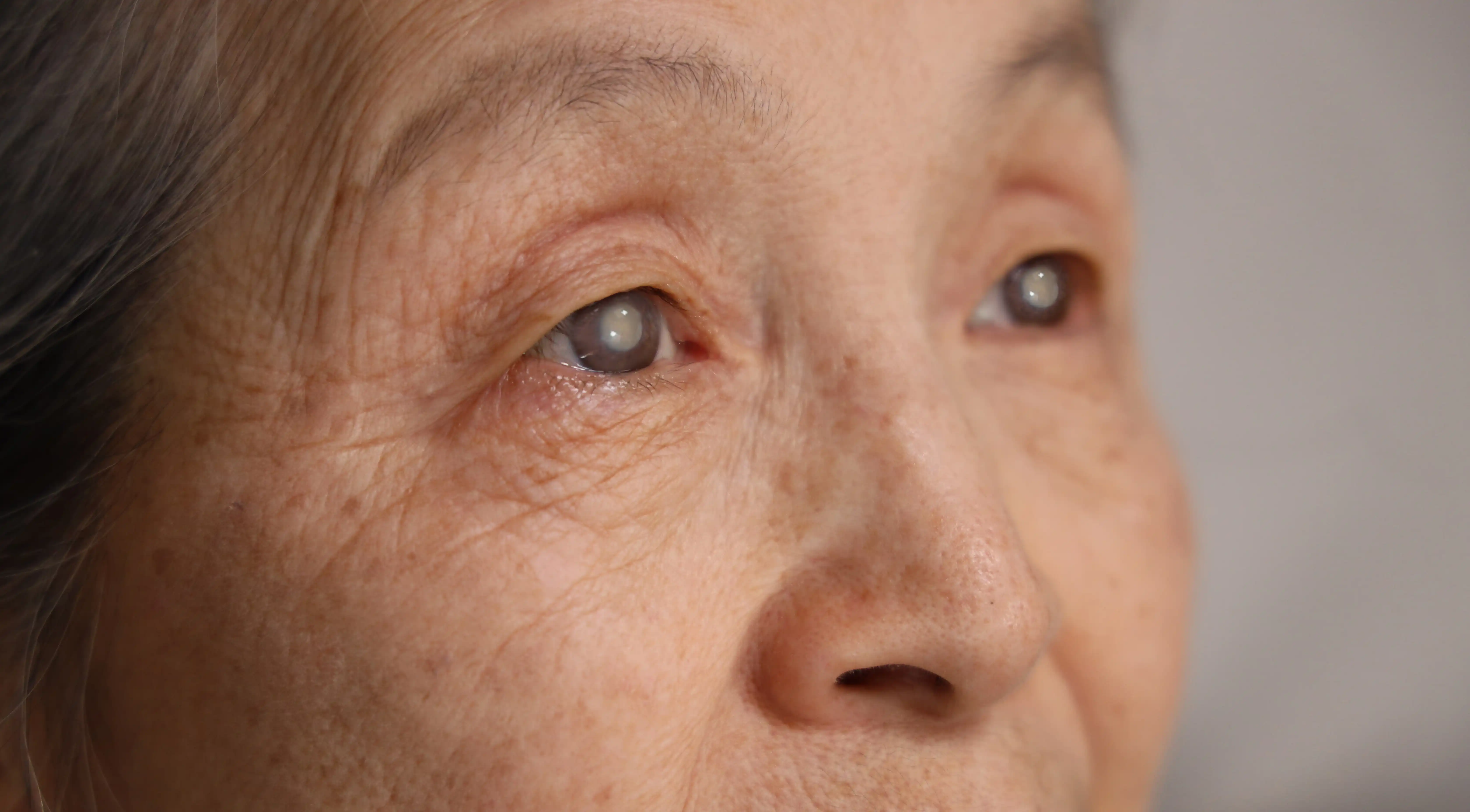
A cataract clouds the natural lens through which light enters the eye. Glaucoma damages the optic nerve. Both can affect vision, and their symptoms may overlap. A patient may describe blur, poor night vision or difficulty reading without being able to identify which condition contributes most.
The surgeon examines the lens, retina, optic nerve and visual field to estimate the likely benefit of cataract removal. The estimate may be less precise if a dense cataract prevents a clear view or reduces test quality. That uncertainty should be discussed before surgery rather than discovered only after the lens has been removed.
Some people with advanced glaucoma still gain useful improvement from cataract surgery. The remaining nerve function, central vision and other eye conditions influence the result. An expectation of improvement is different from a promise of normal vision, and the decision should be connected to the tasks that matter to that particular patient. [4]
Contrast sensitivity is the ability to distinguish an object from its background when the difference between them is small. It helps with tasks such as seeing a curb in dim light or reading faded print. Glaucoma can affect contrast even when high-contrast visual acuity remains relatively good. [2, 3, 5, 6]
This matters when choosing an artificial lens. A design that expands the range of focus may involve optical tradeoffs, and those tradeoffs can matter more in an eye with reduced visual reserve. The consultation should include night driving, glare tolerance, reading needs and the lighting conditions in which the patient struggles.
Two people with the same chart acuity may therefore prefer different lens strategies. One may prioritize sharp distance vision and accept reading glasses. Another with carefully selected early disease may value a wider range of focus. Neither preference should override the limitations imposed by the health of the optic nerve.
An intraocular lens, or IOL, replaces the natural lens removed during cataract surgery. Its power is calculated using measurements of the eye and a planned focusing target. It can correct some refractive error, but it cannot repair the optic nerve or remove an established visual-field defect.
The term premium lens usually refers to particular optical features or pricing arrangements. It is not a guarantee that the lens is medically preferable for every patient. A more expensive option may be a poor match for an eye with advanced disease or a person especially troubled by glare. [7, 8, 9]
Ask the surgeon to describe the expected result with and without glasses at the distances you use most. A lens discussion becomes more useful when it starts with specific tasks, such as reading medication labels, working at a computer or driving at dusk, instead of a broad promise of spectacle freedom.
| Lens type | What it does | With glaucoma |
|---|---|---|
| Monofocal | Sharp focus at one distance, usually far. Glasses for reading | Suits most eyes, including advanced glaucoma [7, 8] |
| Toric | Also corrects regular astigmatism. Can be monofocal | Often suitable when corneal measurements are reliable [14, 18] |
| Enhanced monofocal | Monofocal design with a little more intermediate range | Limited data in glaucoma; ask about the exact lens [10, 11] |
| Extended depth of focus (EDOF) | Wider range of focus, some night halos | Small, short studies, mostly in mild or early glaucoma [27, 28, 83] |
| Multifocal or trifocal | Focus at several distances; more halos and lower contrast | Usually avoided in moderate or advanced glaucoma; selected early, stable cases only [9, 83] |
A standard monofocal IOL is designed primarily for one focusing range. Many patients choose a distance target and use glasses for near work. Others may prefer a different target after discussing their habits and the other eye. The choice of target is separate from the decision to operate.
Some monofocal designs aim to improve intermediate performance while retaining a broadly monofocal approach. Studies of these lenses provide information about visual outcomes, but results in general cataract populations should not automatically be transferred to eyes with substantial glaucoma damage. The exact design and the condition of the eye both matter. [10, 11, 12]
Monovision uses different focusing targets in the two eyes to reduce dependence on glasses for some tasks. It may affect depth perception or binocular comfort and is not suitable for everyone. A history of successful monovision can help the discussion, but a patient should understand the intended compromise before surgery. [13]
A toric IOL corrects a component of regular corneal astigmatism. Toric describes astigmatism correction; it does not necessarily mean multifocal. A lens can be both toric and monofocal. This distinction prevents confusion when a patient is told that glaucoma makes certain optical designs less attractive.
Studies in glaucoma patients, including people undergoing combined cataract and angle surgery, have reported refractive and astigmatism outcomes with toric lenses. Suitability depends on reliable corneal measurements, the type of astigmatism, lens stability and the planned operation. A toric lens cannot correct blur arising from optic nerve damage. [14, 15, 16, 17, 18]
The surgeon also considers whether another planned glaucoma procedure may change the eye's shape or refractive result. Reducing astigmatism can be valuable, but no calculation guarantees that glasses will never be needed. Ask what residual prescription is realistic and how it would be managed. [19]
Multifocal and trifocal lenses distribute light to provide useful focus at more than one distance. They can reduce spectacle dependence in suitable patients, while also producing halos, glare or reductions in some aspects of contrast. Lens designs differ, and results from one design do not apply identically to all others. [20, 21, 22, 23, 24]
Because glaucoma may already reduce contrast and visual-field reserve, these tradeoffs deserve particular attention. More advanced or unstable glaucoma generally makes the choice more difficult. A favorable result in a selected early-disease group is not evidence that the same approach is suitable for an eye with central field loss.
A 2023 systematic review found only 12 studies of premium lenses (toric, multifocal and extended depth of focus) in glaucoma, 399 eyes in all, and some included very few people with glaucoma. They reported good distance vision without glasses and high satisfaction. The reviewers found the best results in people with high eye pressure or early, well-controlled glaucoma, and not enough evidence on safety in advanced glaucoma. They listed the type and severity of field loss, the glaucoma subtype, dry eye, changes from earlier glaucoma surgery and the reliability of future tests as the main things to weigh. [83] A few more small studies have been published since, mostly in mild or early glaucoma.
Research has also examined how multifocal optics influence automated visual-field measurements. A change in a test after implantation may partly reflect the optical system, so future monitoring needs to account for it. Lens selection is therefore relevant not only to everyday vision but also to the interpretation of subsequent tests. [25, 26]
Extended depth of focus, or EDOF, lenses aim to provide a broader useful focusing range. The category includes different optical designs. Describing a lens as EDOF does not by itself specify its contrast performance, night symptoms or suitability for glaucoma. The surgeon should discuss the actual design under consideration.
A prospective study of a non-diffractive EDOF lens included 26 patients with mild open-angle glaucoma and reported favorable distance and intermediate outcomes over a short follow-up period. Many participants also received a trabecular microbypass stent. The study was small and lacked the kind of randomized long-term comparison needed to generalize confidently to more advanced glaucoma. [27]
A separate retrospective comparison in early glaucoma found better intermediate and near performance with the studied EDOF lens than with monofocal lenses, alongside reported differences in spectacle independence. It was nonrandomized and involved a small selected group. These findings support a conversation about carefully selected candidates rather than a universal recommendation. [28]
Other case series and comparative reports add useful experience but do not eliminate uncertainty about future disease progression. A lens that suits the eye today must also be considered in the context of the patient's likely long-term visual needs. [29, 30, 31]
Removing the natural lens can lower pressure in some eyes, but the effect varies with the glaucoma type, starting pressure and anatomy. A cataract operation alone should not be assumed to provide enough pressure reduction for every patient. The surgeon compares the expected effect with the pressure target needed to protect the nerve. [1, 32]
In a 2017 analysis of 32 studies (1,826 people with open-angle glaucoma), pressure was 12% lower than before surgery at six months, 14% at one year, 15% at two years and 9% at three years, and people used slightly fewer drops: about half a drop fewer per person in the first year, falling to 0.16 at three years. [78] A 2024 analysis of the cataract-only groups of nine randomized trials (502 people with open-angle glaucoma or high eye pressure) found pressure 3.8 mmHg lower a year after surgery, and 5.3 mmHg lower in trials that stopped drops before measuring it. [79] In the Ocular Hypertension Treatment Study, 63 eyes with high pressure but no glaucoma, not using pressure drops, had pressure 16.5% lower after cataract surgery, and it stayed lower for at least three years. [80]
Angle-closure disease is a distinct situation because the lens contributes to the crowded anatomy in the front of the eye. Open-angle glaucoma involves a different clinical problem. A treatment result from one group should not be applied indiscriminately to the other. [33, 34, 35]
In the EAGLE trial of people aged 50 or over with angle-closure glaucoma, or angle closure with very high pressure, removing the clear lens worked better than laser iridotomy with drops, and was more cost-effective. The people in EAGLE did not have cataracts, and the gain was modest: pressure about 1.2 mmHg lower and slightly better quality of life after three years. [85] Our guide to angle-closure glaucoma explains when lens removal is offered.
Pressure can also rise temporarily after surgery. Studies have documented early elevations in patients with glaucoma or exfoliation syndrome. The postoperative plan may therefore include specific pressure checks and medication instructions. A longer-term possibility of lower pressure does not remove the need to monitor the immediate recovery period. [36, 37]
In one small randomized study of 122 people having uncomplicated cataract surgery, pressure went above 25 mmHg within the first day in 55% of eyes with well-controlled glaucoma and 27% of eyes with exfoliation syndrome when no pressure-lowering drop was given at the end of surgery, and in none of the eyes without these conditions. Most spikes happened about 4 hours after surgery, and average pressure was below 20 mmHg in every group by the next day. A single drop of timolol at the end of surgery prevented spikes above 30 mmHg, but smaller rises above 25 mmHg still happened in 14% of eyes with glaucoma. [36]
Minimally invasive glaucoma surgery, often called MIGS, includes a range of procedures intended to reduce pressure or medication burden with approaches that differ from traditional filtering surgery. Some are commonly combined with cataract surgery. They are not one interchangeable operation, and not all use an implanted device. [38, 39, 40]
The surgeon should name the proposed procedure and explain its mechanism, expected benefit and limitations. Important questions include whether the drainage angle is suitable, whether the expected reduction meets the pressure target, and what will happen if drops remain necessary. A procedure may reduce medication burden without achieving the very low pressure required in a different clinical situation.
Many angle-based MIGS options are considered in selected patients with open-angle disease. Advanced glaucoma or an especially low target may call for another strategy. The word minimally does not mean risk-free or automatically adequate. The operation must fit the disease rather than being added merely because cataract surgery is already planned.
Randomized studies have compared cataract surgery with and without particular trabecular implants. They help estimate the additional effect of the tested device beyond cataract removal alone. The findings belong to that procedure, trial population and follow-up period; they should not be treated as proof that every MIGS technique produces the same results. [41, 42]
For the Hydrus microstent, a Cochrane review of two trials (about 640 people, mostly mild to moderate glaucoma) found moderate-certainty evidence that adding it to cataract surgery raised the share of people who needed no drops from about half to more than three quarters at one and two years, and, when measured after drops were paused, lowered pressure at two years by about 2 mmHg more than cataract surgery alone. Most people in these trials had pressure that was already controlled on drops. [81] In the HORIZON trial, 66% of eyes that had the stent were off drops at five years, against 46% after cataract surgery alone. [42]
For the iStent, a Cochrane review judged the evidence to be of very low certainty: people who had the iStent with cataract surgery were about 1.4 times as likely to be drop-free at 6 to 18 months, and used about 0.4 of a drop fewer on average. [82] A 2026 analysis of five trials (915 eyes) found much the same, and concluded that the main benefit is fewer drops rather than extra pressure lowering. [88] In the larger trial of the newer iStent inject (505 eyes), pressure measured after drops had been paused for the test was at least 20% lower than at the start in 75.8% of eyes at two years, against 61.9% after cataract surgery alone. [86] All these trials were funded by the device makers.
A five-year post hoc analysis of visual fields from the HORIZON randomized trial found slower average field decline in the group assigned to cataract surgery with a Hydrus microstent than in the cataract-only group. This is relevant evidence about functional outcomes. Because it was a secondary analysis in a defined trial population, it does not establish that all patients or all glaucoma procedures will experience the same benefit. [43]
When discussing a study, ask whether its participants had disease similar to yours and whether the outcome was pressure, medication use, visual-field change or another measure. These outcomes can all matter, but they are not interchangeable. A reduction in drops should be interpreted alongside the continuing need to preserve vision.
Combining procedures may allow the surgeon to address the cataract and pressure control during one operation. A staged approach may be preferable when the pressure problem needs to be addressed first, when refractive planning is particularly complex or when the best sequence depends on the response to an earlier procedure.
Studies comparing combined cataract and filtering operations with other approaches have examined pressure control, complications and refractive outcomes. They do not yield one sequence that is best for every eye. The urgency of glaucoma control, lens opacity, previous surgery and patient circumstances all influence the decision. [44, 45, 46, 47, 48]
Traditional trabeculectomy or a drainage implant may be discussed when the required pressure reduction exceeds what a proposed angle procedure is likely to achieve. These operations have their own risks and follow-up demands. The surgeon should explain why the recommended sequence fits the target rather than present all procedures as equivalent menu choices.
Pressure-lowering surgery may alter aspects of eye shape or measurement, and cataract surgery in an eye with a previous filtering procedure requires attention to that history. Studies have examined changes in axial length, corneal curvature and refractive prediction after trabeculectomy or drainage surgery. [49, 50, 51, 52, 53]
These findings do not mean an accurate result is impossible. They mean the surgeon may need additional records, repeat measurements or a different expectation about refractive precision. The size of a pressure change and the timing between operations can be relevant. [54, 55, 56, 57, 58, 59]
If you have had glaucoma surgery, bring the operative history if the cataract surgeon does not already have it. Ask whether the existing drainage site changes the surgical plan or follow-up. A previous successful procedure still needs to be considered when planning the next one. [60]
A stable tear film helps produce reliable corneal measurements. People using long-term glaucoma drops may also have ocular-surface symptoms that affect comfort or measurement quality. Preparing the surface before final lens calculations can be part of obtaining a dependable plan. [61, 62, 63]
This preparation should be coordinated with the glaucoma regimen. Do not stop pressure-lowering drops simply to make the surface feel better before measurements. Tell the team about burning, fluctuating blur and difficulty tolerating medication so that both pressure control and surface care can be addressed.
A repeated measurement is often a quality step. If readings disagree, the surgeon may want to resolve the cause before committing to a lens power or astigmatism correction. That is especially relevant when a patient has high expectations for vision without glasses.
Pseudoexfoliation can affect the pupil and the structures supporting the natural lens. In some eyes, these factors make cataract surgery more complex and influence the choice or support of the implanted lens. Published studies describe operative difficulties and later lens-related outcomes in this group. [64, 65, 66, 67, 68]
The surgeon may discuss additional devices or techniques if the supporting fibers, called zonules, appear weak. These measures are selected during planning or surgery according to the findings. A general article cannot determine whether they will be needed in an individual eye. Our guide to pseudoexfoliation glaucoma explains the condition.
Other factors, including retinal disease, corneal problems, previous refractive surgery and marked differences between the eyes, can also affect the plan. The glaucoma diagnosis is important, but it should not crowd out assessment of the rest of the visual system.
The National Eye Institute says the surgery lasts about an hour and is almost painless, that most people are completely healed within 8 weeks, and that you may need to avoid touching the eye, bending over and lifting heavy things for a few weeks. You will be given drops to help the eye heal and may need to wear an eye shield. [84] With glaucoma, add an early pressure check, on the day of surgery or the next day depending on how advanced your glaucoma is, and clear instructions about your usual drops.
The postoperative instructions should state which preexisting glaucoma drops to continue, which to pause if instructed, and how to use the new postoperative medicines. Do not assume that successful combined surgery means all glaucoma treatment stops immediately. The plan may change as pressure and healing are assessed.
Steroid drops used after surgery can themselves raise pressure in susceptible patients. Inflammation, retained surgical material and other postoperative factors may also contribute to a pressure change. The surgeon needs to identify the cause rather than have the patient independently reduce the anti-inflammatory treatment.
Ask when pressure will be checked, when activity restrictions will be reviewed and when a new spectacle prescription is likely to be appropriate. Recovery varies by procedure and eye. A fixed timetable found online may not fit a combined operation or a patient with advanced disease.
Before surgery, identify two or three tasks you hope will improve. Examples include seeing faces more clearly, reducing glare from headlights or reading with better lighting and glasses. Ask which goals are realistic given the cataract and which may remain limited by the visual field or optic nerve.
Better central clarity may not restore missing peripheral awareness. A person can be pleased with brighter vision while still needing adaptations for mobility or reading. Low-vision support may remain useful even when the cataract operation is technically successful and the lens is positioned appropriately.
The final spectacle prescription can differ from the calculated target. Studies of cataract and combined glaucoma surgery document this variability. Discuss how residual refractive error would be managed and whether the expected benefit still makes sense if some glasses remain necessary. [69, 70, 71, 72, 73, 74, 75, 76]
Consider an invented example of someone with mild stable open-angle glaucoma, a visually significant cataract and difficulty using several drops. The surgeon may discuss cataract removal with a suitable lens and a particular angle procedure, explaining that fewer drops are possible but not guaranteed. Lens options are considered alongside contrast, night driving and the likelihood of future progression.
Now consider an invented example of someone with advanced central field damage and pressure above the required target. The priority may be dependable pressure control, and the optical tradeoffs of a multifocal design may be less acceptable. A more substantial glaucoma operation or a staged approach may be considered. These examples illustrate the questions, not recommendations for any reader.
The same diagnosis can therefore lead to different plans. The decision should be traceable to the condition of the eye, the pressure target and the person's needs. It should not depend on a general rule that every glaucoma patient must receive one lens or that every cataract operation should include MIGS.
Netra Restoration Therapy (NRT) at Netra Eye Institute does not treat cataracts or glaucoma, does not lower eye pressure, and cannot replace cataract surgery, drops, laser or glaucoma surgery. It does not restore any vision lost to glaucoma. After surgery and once glaucoma is medically stable, some people still struggle with scanning, reading or getting around because of field loss that was already there. Low-vision optometrists and occupational therapists can help with those tasks. If you choose NRT, it is offered only alongside that care and your glaucoma treatment, never in place of them.
Learn about Netra Restoration Therapy and low-vision rehabilitation.
Yes. Most people with glaucoma can have cataract surgery, and some have it at the same time as a glaucoma procedure. The surgeon first checks that the pressure is controlled well enough, looks at the drainage angle and optic nerve, and plans the lens and follow-up around your glaucoma. Advanced glaucoma or very high pressure may change the order of operations.
Often a little. In people with open-angle glaucoma, pressure was about 12% to 15% lower in the first two years and 9% lower at three years across 32 studies, and people used slightly fewer drops. In angle-closure glaucoma, removing the lens can widen the drainage angle, and the fall is usually larger. The effect varies, so your glaucoma doctor keeps measuring pressure after surgery.
It is worth asking about if you have mild to moderate open-angle glaucoma, especially if you use several drops. Adding a Hydrus microstent increased the share of people who needed no drops from about half to more than three quarters in two trials; evidence for the iStent is weaker. Most MIGS procedures lower pressure less than trabeculectomy or a tube, so they may not be enough for advanced glaucoma. [38, 39]
There is no single best lens, but a monofocal lens, with toric correction for astigmatism if needed, suits most eyes with glaucoma. Multifocal and extended depth of focus lenses can reduce the need for glasses but may lower contrast and cause halos, which matters more when glaucoma has already reduced contrast or side vision. They are usually kept for selected people with early, stable glaucoma.
Not on your own. Some people need fewer drops afterward, but pressure can rise in the first days, and the steroid drops used after surgery can raise it too. Keep using your glaucoma drops exactly as your surgeon instructs, and let your glaucoma doctor decide whether any can be reduced once the eye has healed.
Glaucoma adds a few specific risks: a pressure spike in the first hours, a pressure rise from steroid drops, and in eyes with pseudoexfoliation, a small pupil and weak lens supports that make surgery harder. A previous trabeculectomy also needs planning, because cataract surgery can make it drain less well in some eyes. [60] These risks are usually managed with extra pressure checks and surgical planning, and serious complications remain uncommon.
Many people see more clearly within a few days, although vision can be blurry at first, and most are completely healed within about 8 weeks. Expect an early pressure check, on the day of surgery or the next day, then more visits than after cataract surgery alone, especially if a glaucoma procedure was added. A new glasses prescription usually waits until the eye has settled.
It can raise eye pressure for a while, from inflammation, leftover gel used during surgery or a response to steroid drops, and this usually settles with treatment. Lasting glaucoma after adult cataract surgery is not common. Children are different: across 36 studies, about 7% developed glaucoma after surgery for cataracts they were born with, and about 14% of those left without a lens implant, so they need lifelong pressure checks. [89] Our guide to glaucoma vs cataracts covers this in more detail.
No. It may improve cataract-related vision and can affect pressure, but glaucoma still requires follow-up. Even when medication burden falls, the optic nerve and visual field need continued assessment. Existing nerve damage is not removed along with the cloudy lens.
No universal statement fits every design and every stage of glaucoma. Selected patients with early stable disease may have options that would be less appropriate in advanced disease. Ask about the evidence for the exact lens and the tradeoffs relevant to your remaining contrast and field function.
No. Some patients reduce medication use, while others continue drops or need additional treatment later. The intended goal should be explained before surgery. A good result may be improved pressure control on fewer medicines, rather than permanent freedom from every treatment.
The clinician may establish new postoperative imaging or field baselines after the eye has recovered, because removing a cataract and changing the optical system can affect test quality or measurements. The timing and interpretation depend on the individual record. An apparent improvement in a field index does not mean lost nerve tissue regrew. [25, 26, 77]
Contact the surgical team urgently for increasing pain, marked redness, sudden or worsening vision loss, new flashes or a curtain-like shadow, severe eye pain or headache with nausea or vomiting, rainbow-colored halos around lights, or other symptoms identified in your discharge instructions. Do not assume that a major change is normal recovery or simply the glaucoma. If the surgical service cannot be reached, seek urgent ophthalmic care.
The most useful preoperative plan states the expected visual benefit, the lens target, the pressure-control strategy and the follow-up responsibilities. With glaucoma, each part supports the others. Clear expectations help patients judge the outcome by meaningful improvements while continuing the care needed to protect their remaining vision.
Numbered citations identify sources connected to the text. The remaining entries provide relevant supporting research. Clinical trials, observational studies, laboratory research and professional guidance answer different questions; the size of this bibliography does not imply that every source provides equally strong evidence.
Medical Disclaimer: This article provides general education. It is not medical advice, a diagnosis or a personal treatment plan, and it does not establish a doctor-patient relationship. Do not start, stop or change any prescribed medicine or treatment without the clinicians responsible for your care. Sudden vision loss, a new shower of floaters, flashes, a curtain or shadow in your vision, or a painful red eye needs urgent eye care.