
Blog
Ocular Blood Flow and Vascular Dysregulation in Glaucoma
June 28, 2026
Key takeaways on ocular blood flow and glaucoma
- Glaucoma is a progressive optic neuropathy in which retinal ganglion cells and their axons die, causing irreversible vision loss.
- High intraocular pressure (eye pressure) is the leading treatable risk factor, but it does not fully explain why many patients progress.
- Reduced and unstable ocular blood flow is consistently observed in glaucoma, particularly normal-tension glaucoma.
- Vascular dysregulation is the failure of small ocular vessels to keep blood flow steady when pressure, posture, or temperature change.
- Faulty autoregulation at the optic nerve head exposes axons to cycles of low oxygen (ischemia) and reperfusion injury.
- Nighttime blood pressure dipping and systemic hypotension are linked to faster visual field loss in several cohort studies.
- Endothelial dysfunction, reduced nitric oxide, and raised endothelin-1 are mechanisms tied to abnormal ocular perfusion.
- Primary vascular dysregulation, marked by cold extremities, low blood pressure, and migraine, is more common in normal-tension glaucoma.
- Optical coherence tomography angiography (OCTA) now lets clinicians measure peripapillary and macular vessel density non-invasively.
- Lowering eye pressure remains first-line care; vascular strategies are adjunctive and partly still under investigation.
- Emerging therapies target neuroprotection, blood flow, and mitochondrial health, but most have moderate or preliminary evidence only.
Why does glaucoma keep getting worse when eye pressure is normal?
Because eye pressure is only one of the forces acting on the optic nerve, and blood supply is another. Glaucoma affects roughly 80 million people worldwide and is a leading cause of irreversible blindness. Lowering intraocular pressure slows it in most patients, yet a meaningful minority continue to lose vision despite "good" numbers.
The vascular theory of glaucoma helps fill that gap. The optic nerve head is metabolically hungry and depends on a steady, well-regulated blood supply. When that supply becomes unstable, nerve fibers suffer even at normal pressure. This is why understanding ocular blood flow and vascular dysregulation matters for anyone trying to protect sight over decades.
This article explains the normal blood supply of the optic nerve, what goes wrong in vascular dysregulation, how clinicians measure it, and how treatment is evolving. It is written for patients, optometry and medical students, practising eye doctors, and researchers at once. You can read more about the broader disease on our glaucoma overview page.
What is glaucoma and how does blood flow fit in?
Glaucoma is a group of optic neuropathies defined by progressive loss of retinal ganglion cells, thinning of the retinal nerve fiber layer, and characteristic cupping of the optic nerve head, leading to corresponding visual field loss. The classic risk factor is elevated intraocular pressure, but the disease is now understood as multifactorial, with mechanical and vascular components.
The vascular contribution was proposed over a century ago and revived through detailed studies of optic nerve head perfusion. Researchers such as Josef Flammer described how poor ocular blood flow regulation, not just pressure, can injure the nerve. This thinking is most relevant in normal-tension glaucoma, where damage occurs at statistically normal pressures.
Key Point: Eye pressure and blood flow are not competing explanations. In most patients they act together, which is why controlling pressure helps but does not always stop progression.
| Quick fact | Detail |
|---|---|
| Global cases | Roughly 80 million people, projected to rise with aging populations |
| Main treatable risk factor | Elevated intraocular pressure (eye pressure) |
| Vascular-prominent subtype | Normal-tension glaucoma |
| Cells lost | Retinal ganglion cells and their axons |
| Reversibility | Vision lost is permanent; the goal is to slow further loss |
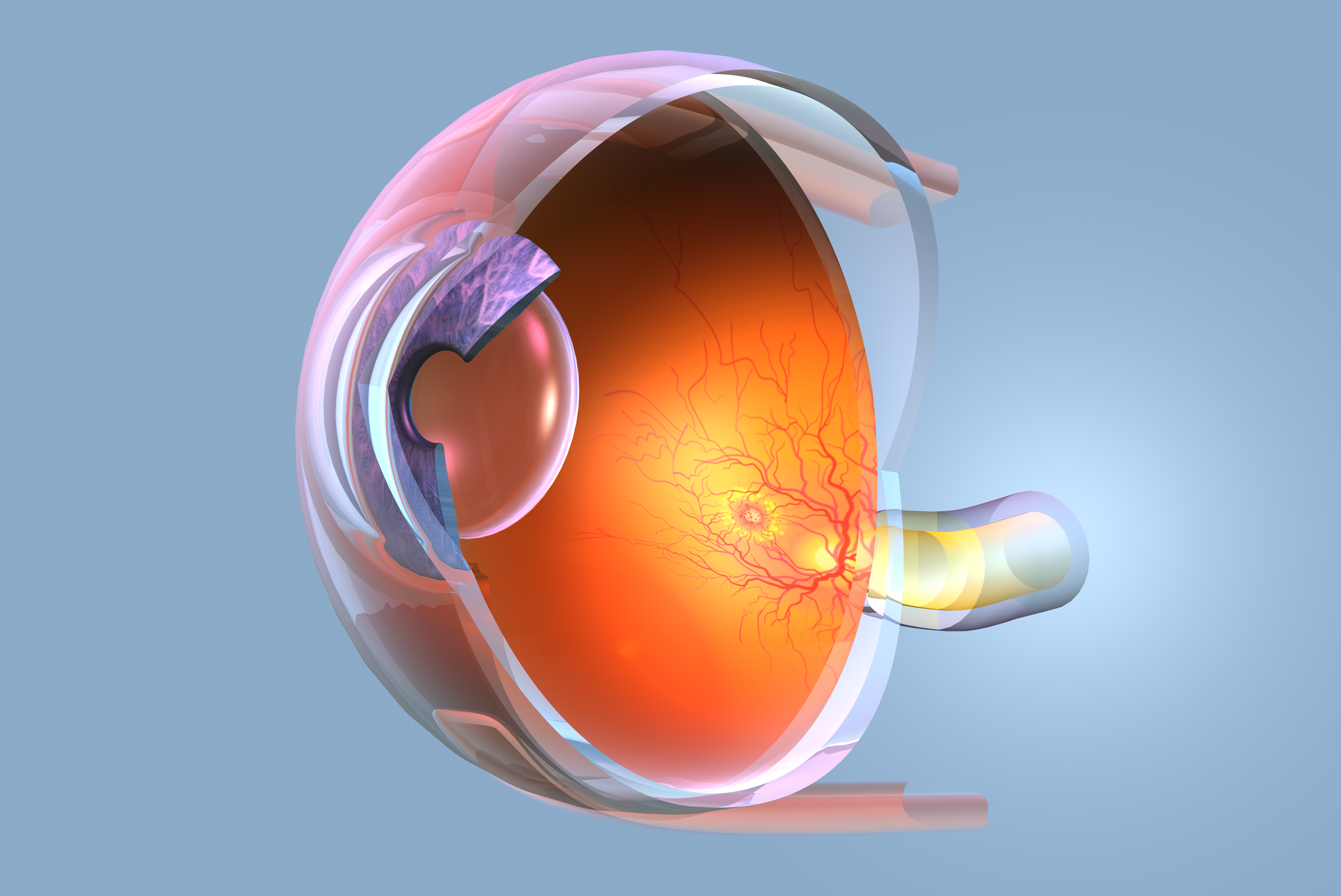
How does blood normally reach the optic nerve?
The optic nerve head receives blood mainly from the posterior ciliary arteries, branches of the ophthalmic artery, which itself arises from the internal carotid artery. The retina is fed by the central retinal artery, while the deeper optic nerve head and choroid depend heavily on the short posterior ciliary arteries and the circle of Zinn-Haller.
This blood supply is unusual because it sits in a pressure-sensitive environment. Perfusion pressure at the eye equals blood pressure minus intraocular pressure. So when eye pressure rises or blood pressure falls, the pressure pushing blood through the nerve drops. Healthy vessels compensate through autoregulation, adjusting their diameter to keep flow constant.
What is ocular autoregulation and why does it matter?
Autoregulation is the ability of ocular vessels to maintain steady blood flow despite changes in perfusion pressure. The retina and optic nerve head have strong autoregulatory capacity, controlled by local factors including nitric oxide, endothelin-1, carbon dioxide, and metabolic demand, rather than by autonomic nerves alone.
When autoregulation works, a brief rise in eye pressure or a drop in blood pressure does little harm. When it fails, the nerve is exposed directly to swings in perfusion. This is the central idea behind vascular dysregulation in glaucoma.
How do tiny vessels in the eye keep flow steady?
Ocular microvessels rely on the endothelium, the inner lining of blood vessels, which releases nitric oxide to relax vessels and endothelin-1 to constrict them. Pericytes wrapped around capillaries also fine-tune flow. Healthy balance keeps oxygen delivery matched to the high metabolic needs of nerve axons and mitochondria.
When this balance tips toward constriction or becomes erratic, the optic nerve head experiences fluctuating oxygen levels. These swings, rather than a single dramatic blockage, are thought to drive slow glaucomatous damage.
What are the signs and symptoms across glaucoma stages?
Glaucoma is usually painless and silent until vision is already lost, which is why it is called the silent thief of sight. Vascular factors can make progression faster or more erratic, but the symptoms by stage are similar to glaucoma overall.
| Stage | What is happening | What the patient notices |
|---|---|---|
| Early | Mild nerve fiber loss, small visual field defects | Usually nothing; detected on testing |
| Intermediate | Expanding field defects, more cupping | Occasional missed objects, mild difficulty in dim light |
| Advanced | Significant field loss, central vision threatened | Bumping into things, tunnel-like vision |
| Emergency | Acute angle-closure or sudden pressure spike | Severe eye pain, redness, halos, nausea, sudden blurring |
Clinical Pearl: Patients with strong vascular features may report cold hands and feet, low blood pressure, or migraine. These clues raise suspicion for normal-tension glaucoma and prompt closer blood pressure review.
Acute angle-closure with severe pain and nausea is a true emergency. If you have sudden, severe eye pain with halos and vomiting, seek urgent care. Learn more on our angle-closure glaucoma page.
What causes vascular dysregulation in glaucoma?
Vascular dysregulation arises from a mix of systemic vascular tendencies, local endothelial problems, and blood pressure behaviour. Some causes are fixed, others can be modified.
Primary vascular dysregulation: who has it?
Primary vascular dysregulation is a constitutional tendency for blood vessels to respond abnormally to stimuli such as cold or stress. People with it often have cold hands and feet, low or borderline blood pressure, a tendency to faint, and migraine. It is more common in women, in slender individuals, and in patients with normal-tension glaucoma.
Secondary causes that disturb ocular perfusion
Secondary vascular dysregulation follows other conditions such as autoimmune disease, sleep apnea, atherosclerosis, or carotid disease. Obstructive sleep apnea causes repeated nighttime drops in oxygen and surges in blood pressure that stress the optic nerve circulation.
| Non-modifiable factors | Modifiable factors |
|---|---|
| Age | Untreated obstructive sleep apnea |
| Family history | Aggressive nighttime blood pressure lowering |
| Female sex (for primary vascular dysregulation) | Smoking |
| Genetic predisposition | Dehydration and poor cardiovascular fitness |
| Ethnic background (varies by glaucoma type) | Untreated systemic hypotension or hypertension |
What is the pathophysiology of vascular damage in glaucoma?
Vascular dysregulation injures the optic nerve through repeated mismatches between blood supply and the high oxygen demand of axons. Several mechanisms work together; each is described below as a standalone process.
How does unstable ocular perfusion pressure harm the nerve?
Ocular perfusion pressure is blood pressure minus intraocular pressure. When this number swings, the optic nerve head experiences alternating periods of adequate and inadequate blood flow. The Barbados Eye Studies and other population research linked low ocular perfusion pressure to higher glaucoma risk. Unstable perfusion appears more damaging than a steady, modestly reduced level, because fluctuation drives ischemia-reperfusion stress.
Why is nighttime blood pressure dipping a problem?
Blood pressure naturally falls during sleep, but in some people it falls too far, a pattern called over-dipping. When blood pressure plunges at night while a person lies flat, optic nerve perfusion can drop below what damaged autoregulation can buffer. Several observational studies have associated nocturnal hypotension and over-dipping with faster visual field progression, especially in normal-tension glaucoma. This is why aggressive evening dosing of blood pressure drugs can sometimes backfire for the eye.
Evidence Snapshot: Observational cohorts link nocturnal blood pressure dips to faster field loss, but randomized trials proving that adjusting medication timing protects vision are limited. Treat this as a plausible, evidence-supported concern rather than settled fact.
What role does endothelial dysfunction play?
Endothelial dysfunction means the vessel lining fails to balance dilation and constriction properly. In glaucoma, studies report reduced nitric oxide availability and elevated endothelin-1, a powerful vasoconstrictor. Higher endothelin-1 levels have been measured in the blood and aqueous humor of some glaucoma patients. The result is a tendency toward vasoconstriction and impaired flow reserve at the optic nerve head.
How does ischemia-reperfusion injure ganglion cells?
When blood flow briefly drops then returns, the reperfusion phase floods tissue with oxygen and generates reactive oxygen species. These free radicals damage mitochondria, proteins, and membranes. Retinal ganglion cell axons, which are densely packed with energy-hungry mitochondria at the optic nerve head, are particularly vulnerable to this oxidative stress.
Why do mitochondria matter in vascular glaucoma?
Mitochondria supply the energy that ganglion cells need to maintain their long axons and electrical signaling. Repeated low-oxygen episodes impair mitochondrial function, lower ATP production, and trigger pathways leading to apoptosis, a form of programmed cell death. This links the vascular theory to the broader idea of glaucoma as a metabolic and neurodegenerative disease.
Does the blood-retinal barrier and neurovascular unit break down?
The neurovascular unit is the close partnership between neurons, glial cells, and blood vessels that matches blood flow to activity. In glaucoma, dysfunction of this unit and of the blood-retinal barrier may worsen flow control and allow harmful molecules into delicate tissue. This is an active area of research with mostly preclinical and early human evidence.
How do doctors diagnose vascular involvement in glaucoma?
Diagnosis combines standard glaucoma assessment with tools that probe blood flow and blood pressure behaviour. There is no single test that proves vascular dysregulation, so clinicians build a picture from several sources.
Core glaucoma evaluation includes intraocular pressure measurement, gonioscopy, optic nerve examination, optical coherence tomography (OCT) of the nerve fiber layer, and visual field testing. Vascular evaluation adds blood flow imaging, blood pressure profiling, and a careful history of cold extremities, migraine, and fainting.
| Test | What it measures | Vascular relevance |
|---|---|---|
| OCT angiography (OCTA) | Peripapillary and macular vessel density | Reduced vessel density correlates with damage |
| 24-hour blood pressure monitoring | Daytime and nighttime blood pressure | Detects over-dipping and nocturnal hypotension |
| Visual field testing | Functional field loss over time | Tracks progression that may be vascular |
| OCT (RNFL and GCC) | Structural nerve fiber and ganglion cell layer | Baseline for structure-function correlation |
| Nailfold capillaroscopy (research) | Microvascular reactivity to cold | Supports primary vascular dysregulation |
Research Update: OCT angiography has made non-invasive measurement of optic nerve and macular perfusion routine. Studies consistently show lower peripapillary vessel density in glaucoma, though whether reduced flow is fully cause or partly consequence is still debated.
For more on imaging, see our OCT and OCT angiography explainer.
How is glaucoma treated, and where does blood flow fit in?
Lowering intraocular pressure is the only proven, first-line way to slow glaucoma, and it remains the backbone of care regardless of vascular features. Vascular management is adjunctive and aims to stabilise perfusion rather than replace pressure-lowering.
Medical, laser, and surgical pressure-lowering
Pressure can be lowered with eye drops, laser trabeculoplasty, or surgery. Each route reduces the mechanical and indirectly the perfusion stress on the optic nerve, since lower eye pressure raises ocular perfusion pressure.
| Treatment | How it helps | Limitations |
|---|---|---|
| Prostaglandin analog drops | Strong pressure lowering, once daily | Do not directly target blood flow |
| Beta-blocker drops | Lower pressure | Can lower systemic and nighttime blood pressure, a vascular concern |
| Selective laser trabeculoplasty | Lowers pressure, reduces drop burden | Effect may wane over years |
| Filtering or MIGS surgery | Substantial pressure lowering | Surgical risks; no direct blood flow effect |
Clinical Pearl: In patients with low blood pressure and suspected vascular dysregulation, clinicians may avoid systemic beta-blocker effects and review the timing of any blood pressure medication with the patient's physician.
Why do some patients still lose vision despite good treatment?
Some patients progress because pressure control alone does not correct unstable blood flow, ongoing oxidative stress, or systemic vascular problems. The landmark Collaborative Normal-Tension Glaucoma Study showed that lowering pressure slows progression even at normal pressures, yet a subset of treated eyes still worsened, pointing to non-pressure factors.
Contributors to continued loss include nocturnal hypotension, sleep apnea, persistent vasospasm, advanced baseline damage, and individual genetic vulnerability. Recognising these helps target the right additional strategies, such as treating sleep apnea or coordinating blood pressure care, rather than simply pushing eye pressure ever lower.
What emerging therapies target blood flow and nerve survival?
Emerging therapies aim to protect ganglion cells (neuroprotection), improve or stabilise blood flow, and support mitochondria. Evidence ranges from preclinical to moderate; none replaces pressure-lowering today.
| Approach | Goal | Evidence level |
|---|---|---|
| Calcium channel blockers (low dose) | Reduce vasospasm, improve ocular flow | Mixed, small human studies |
| Ginkgo biloba extract | Improve microcirculation, antioxidant effect | Emerging, small trials |
| Nicotinamide (vitamin B3) | Support mitochondrial energy in ganglion cells | Promising early human and animal data |
| Citicoline | Neuroprotection of visual pathway | Moderate, several small trials |
| Gene and stem cell therapy | Protect or replace ganglion cells | Preclinical to early trial |
| Photobiomodulation | Support mitochondrial function | Preclinical and limited human data |
Evidence Snapshot: A 2017 crossover study of nicotinamide in glaucoma reported improved inner retinal function, and a 2022 trial suggested benefit alongside pressure-lowering. These results are encouraging but need larger, longer trials before nicotinamide becomes standard.
Patients should not start supplements alone for glaucoma. Discuss any of these with your eye doctor, and see our glaucoma neuroprotection resource.
What does an integrative ophthalmology perspective add?
An integrative perspective treats glaucoma as a whole-body vascular and metabolic problem, using lifestyle and systemic measures to support, never replace, standard eye care. The mechanisms it targets are real: blood pressure stability, vascular health, oxidative stress, and stress physiology.
- Cardiovascular fitness: Regular aerobic exercise supports healthy endothelial function and can modestly lower eye pressure.
- Avoiding nocturnal hypotension: Coordinating blood pressure medication timing with a physician may protect optic nerve perfusion.
- Sleep apnea treatment: Treating obstructive sleep apnea reduces nighttime oxygen swings.
- Nutrition: Diets rich in leafy greens and nitrate-containing vegetables may support nitric oxide and vascular health, based on observational data.
- Stress and autonomic balance: Chronic stress affects vascular tone; relaxation practices may help, though direct glaucoma evidence is limited.
Some traditional systems such as Traditional Chinese Medicine and Ayurveda emphasise circulation and stress, and certain herbal compounds are being studied for antioxidant and vasoactive effects. Evidence remains early, and these should be viewed as complementary, not curative.
How does Netra Restoration Therapy approach ocular blood flow?
Netra Restoration Therapy is an integrative, adjunctive approach intended to complement, not replace, standard glaucoma treatment, with proposed mechanisms centered on ocular blood flow and nerve health. Research in this area continues to evolve, and no specific visual outcome can be promised.
The proposed mechanisms map onto the vascular pathways described in this article:
- Ocular blood flow support: Aiming to stabilise perfusion to the optic nerve head.
- Oxidative stress reduction: Limiting free radical damage from ischemia-reperfusion cycles.
- Inflammatory regulation: Calming low-grade inflammation that can affect vessels.
- Mitochondrial and neurotrophic support: Helping ganglion cells maintain energy and survival signals.
- Autonomic balance: Supporting steady vascular tone and blood pressure regulation.
Key Point: Any integrative therapy, including Netra Restoration Therapy, should be used alongside prescribed pressure-lowering treatment and regular monitoring. Continue your standard glaucoma care and keep all follow-up appointments.
What does the clinical evidence say so far?
The evidence base mixes strong epidemiology linking blood flow to glaucoma with smaller, less conclusive treatment trials. The table below summarises representative findings in one honest line each.
| Study or body of work | Type | Key finding (one line) |
|---|---|---|
| Collaborative Normal-Tension Glaucoma Study | Randomized trial | Lowering pressure slowed progression at normal pressures, but some eyes still worsened. |
| Barbados Eye Studies | Population cohort | Low ocular perfusion pressure associated with higher open-angle glaucoma risk. |
| Egna-Neumarkt Study | Population cohort | Linked low diastolic perfusion pressure to greater glaucoma prevalence. |
| OCTA vessel density studies | Cross-sectional imaging | Reduced peripapillary vessel density correlates with structural and functional loss. |
| Nicotinamide trials (2017, 2022) | Small clinical trials | Suggested improved retinal function as add-on therapy; larger trials needed. |
What are common myths about blood flow and glaucoma?
| Myth | Fact |
|---|---|
| Normal eye pressure means no glaucoma risk | Normal-tension glaucoma damages the nerve at statistically normal pressures. |
| Only eye pressure matters | Blood flow, blood pressure, and oxidative stress also influence progression. |
| Higher blood pressure is always good for the eye | Both very low and very high blood pressure can harm optic nerve perfusion. |
| Supplements can replace eye drops | No supplement replaces proven pressure-lowering treatment. |
| Glaucoma vision loss can be reversed with better circulation | Lost vision is permanent; the goal is to slow further loss. |
Frequently asked questions about ocular blood flow and glaucoma
Can glaucoma progress even with normal eye pressure?
Yes. Normal-tension glaucoma damages the optic nerve at statistically normal pressures, and reduced or unstable ocular blood flow is a leading explanation. Lowering pressure further still helps many of these patients, but vascular factors such as nighttime blood pressure dips and vasospasm can drive progression independently. Careful monitoring and attention to systemic vascular health are important.
What is vascular dysregulation in simple terms?
Vascular dysregulation means the small blood vessels in your eye and body do not keep blood flow steady the way they should. They may tighten too much, fail to relax when needed, or overreact to cold and stress. In glaucoma, this leads to repeated brief shortages of oxygen to the optic nerve, which can slowly damage it over time.
Does low blood pressure cause glaucoma?
Low blood pressure does not directly cause glaucoma, but it can contribute to progression by lowering the pressure that drives blood through the optic nerve. Nighttime drops are especially relevant. If your blood pressure runs low or dips sharply at night, your eye doctor may coordinate with your physician about medication timing, but you should never change prescriptions on your own.
Should I stop my blood pressure medication to protect my eyes?
No. Stopping blood pressure medication on your own can be dangerous and raise your risk of stroke and heart disease. If you and your eye doctor suspect nighttime over-dipping is harming your optic nerve, the right step is a conversation with the physician who manages your blood pressure about dosing and timing, supported by 24-hour blood pressure monitoring.
What is ocular perfusion pressure?
Ocular perfusion pressure is roughly your blood pressure minus your eye pressure. It represents the force available to push blood through the optic nerve and retina. When eye pressure rises or blood pressure falls, perfusion pressure drops. Studies link low ocular perfusion pressure to higher glaucoma risk, which is why both numbers matter together rather than in isolation.
Can exercise help my glaucoma?
Regular aerobic exercise supports healthy blood vessels and can modestly lower eye pressure in many people. It also improves overall cardiovascular health, which benefits optic nerve perfusion. Some high-resistance activities and certain yoga inversions can briefly raise eye pressure, so ask your eye doctor for guidance. Exercise complements, but does not replace, your prescribed treatment.
Does cold sensitivity relate to my glaucoma?
It can. Cold hands and feet, low blood pressure, and migraine are signs of primary vascular dysregulation, a tendency for blood vessels to overreact. This pattern is more common in people with normal-tension glaucoma. Mentioning these symptoms to your eye doctor helps them assess the vascular side of your disease and consider appropriate monitoring.
Is normal-tension glaucoma more vascular than other types?
Vascular factors appear especially important in normal-tension glaucoma, where damage occurs without high eye pressure. Patients more often show signs of vascular dysregulation, nighttime hypotension, and migraine. That said, vascular contributions also matter in high-pressure glaucoma. The difference is one of emphasis, not an absolute divide between types.
Can OCT angiography diagnose vascular glaucoma?
OCT angiography measures blood vessel density in the retina and around the optic nerve without injections. It consistently shows reduced vessel density in glaucoma and helps track damage. However, it cannot by itself prove that poor blood flow caused the disease, since reduced flow can be partly a consequence of nerve loss. It is one valuable piece of the assessment.
Will improving circulation reverse my vision loss?
No. Vision lost to glaucoma is permanent because dead retinal ganglion cells do not regrow with current treatments. The realistic goal is to slow or stop further loss. Supporting healthy circulation and stable blood pressure may help protect remaining nerve fibers, but it works alongside pressure-lowering treatment rather than restoring sight already lost.
Are supplements like ginkgo or nicotinamide proven to work?
Not yet proven, but promising for some. Small studies of ginkgo biloba and nicotinamide (vitamin B3) suggest possible benefits for blood flow and nerve energy, and larger trials are underway. Because supplements can interact with medications and quality varies, discuss any product with your eye doctor before starting it, and never use it to replace prescribed treatment.
Does sleep apnea affect glaucoma?
Yes. Obstructive sleep apnea causes repeated drops in blood oxygen and surges in blood pressure during sleep, which stress the optic nerve circulation. Untreated sleep apnea is associated with higher glaucoma risk and possibly faster progression. If you snore heavily, wake gasping, or feel unrefreshed, ask about a sleep study; treating apnea benefits both your eyes and your heart.
How often should I be monitored if I have vascular features?
That depends on your damage and progression rate, but patients with vascular features and normal-tension glaucoma often need careful, regular follow-up with visual fields and OCT. Your eye doctor sets the schedule based on how stable you are. Consistent monitoring catches progression early, when changing treatment can still protect remaining vision.
Can stress make glaucoma worse?
Chronic stress affects blood pressure and vascular tone, which could in theory influence optic nerve perfusion, though direct evidence in glaucoma is limited. Managing stress supports overall vascular health and wellbeing. Relaxation techniques are reasonable as part of a healthy lifestyle but should accompany, not replace, your prescribed glaucoma treatment and monitoring.
Is glaucoma a neurodegenerative disease?
Increasingly, yes. Glaucoma shares features with other neurodegenerative conditions, including mitochondrial dysfunction, oxidative stress, and progressive neuron loss. The vascular theory fits this view, since unstable blood flow contributes to the metabolic stress that kills retinal ganglion cells. This broader understanding is shaping new research into neuroprotection.
Does caffeine raise my eye pressure?
Caffeine can cause a small, short-lived rise in eye pressure in some people, but moderate consumption is generally considered acceptable for most patients. Very high intake may have a larger effect. If you have glaucoma, discuss your usual caffeine habits with your eye doctor rather than making drastic changes on your own.
Can dehydration affect my eyes and glaucoma?
Severe dehydration can lower blood pressure and reduce perfusion, which is not ideal for the optic nerve, though drinking a very large volume of fluid quickly can briefly raise eye pressure. Sensible, steady hydration through the day is the practical advice. As always, fit this into the broader plan your eye doctor recommends.
What is endothelin-1 and why does it matter?
Endothelin-1 is a powerful molecule made by blood vessel walls that causes vessels to constrict. Some glaucoma patients have higher levels, which may reduce blood flow to the optic nerve. Researchers are studying whether blocking endothelin-1 could protect vision. For now it is an important mechanism in the vascular theory rather than a treatment target in clinic.
Can young people get vascular glaucoma?
Glaucoma is more common with age, but younger people, particularly slim women with low blood pressure, cold extremities, and migraine, can develop normal-tension glaucoma with strong vascular features. Family history raises risk. Anyone with these patterns and a family history of glaucoma should have a thorough eye examination including optic nerve assessment.
Does lowering eye pressure improve blood flow too?
Yes, indirectly. Because ocular perfusion pressure equals blood pressure minus eye pressure, lowering eye pressure raises the pressure available to drive blood through the optic nerve. This is one reason pressure-lowering helps even patients whose problem is partly vascular. It does not fix faulty autoregulation, but it relieves some of the strain.
Are there glaucoma drops that improve blood flow?
Some medications have been studied for blood flow effects beyond pressure lowering. Certain agents may improve optic nerve perfusion, while others, like systemic beta-blocker effects, can lower nighttime blood pressure unhelpfully. Your eye doctor weighs these factors when choosing drops, especially if you have low blood pressure or vascular dysregulation.
Should I monitor my blood pressure at home?
Home or 24-hour blood pressure monitoring can be very useful if vascular factors are suspected, because it reveals nighttime dips that clinic readings miss. Share the results with both your eye doctor and the physician who manages your blood pressure so they can coordinate care. Do not adjust any medication based on home readings without medical advice.
Can glaucoma damage be slowed if caught early?
Yes. Early detection and treatment are the most powerful tools against glaucoma. The earlier pressure is controlled and any vascular contributors are addressed, the more nerve tissue you can preserve. Because early glaucoma has no symptoms, regular eye exams, especially if you have risk factors, are the key to catching it in time.
Is glaucoma hereditary?
Family history significantly increases glaucoma risk, including normal-tension and vascular-prominent forms. If a close relative has glaucoma, your risk is several times higher, and you should have regular comprehensive eye exams. Genetics interact with vascular and other factors, so a family history is a reason for vigilance rather than a guarantee of disease.
Does smoking harm the optic nerve circulation?
Yes. Smoking damages blood vessels, promotes endothelial dysfunction, and increases oxidative stress, all of which are harmful to optic nerve perfusion. Quitting smoking benefits your eyes along with your heart, lungs, and brain. It is one of the most impactful modifiable steps you can take to support your overall and ocular vascular health.
Glossary of key terms
- Intraocular pressure (IOP): The fluid pressure inside the eye, the main treatable glaucoma risk factor.
- Ocular perfusion pressure: Blood pressure minus eye pressure; the force driving blood through the eye.
- Autoregulation: The vessels' ability to keep blood flow steady despite pressure changes.
- Vascular dysregulation: Faulty control of blood vessel diameter and flow.
- Endothelium: The inner lining of blood vessels that controls dilation and constriction.
- Endothelin-1: A strong vessel-constricting molecule made by the endothelium.
- Nitric oxide: A molecule that relaxes blood vessels and improves flow.
- Ischemia-reperfusion: Damage from blood flow dropping then returning, generating free radicals.
- Retinal ganglion cells: The neurons whose axons form the optic nerve and die in glaucoma.
- Normal-tension glaucoma: Glaucoma occurring at statistically normal eye pressures.
- OCT angiography (OCTA): Non-invasive imaging of retinal and optic nerve blood vessels.
- Nocturnal dipping: The natural fall in blood pressure during sleep; over-dipping is excessive.
Summary
Glaucoma is a progressive optic neuropathy in which lowering intraocular pressure slows but does not always stop vision loss. Ocular blood flow and vascular dysregulation help explain continued progression, especially in normal-tension glaucoma. The optic nerve depends on steady, well-regulated perfusion, and when autoregulation fails, the nerve suffers repeated cycles of ischemia and reperfusion, oxidative stress, and mitochondrial strain.
Diagnosis blends standard glaucoma testing with OCT angiography and blood pressure profiling. Treatment still centers on pressure-lowering, while vascular strategies, including managing blood pressure timing, treating sleep apnea, and supporting cardiovascular health, are adjunctive. Emerging therapies and integrative approaches, including Netra Restoration Therapy, target blood flow, oxidative stress, and nerve survival, but they complement standard care and promise nothing. Early detection and consistent monitoring remain the strongest tools for protecting sight.
References
- Flammer J, Orgül S, Costa VP, et al. The impact of ocular blood flow in glaucoma. Prog Retin Eye Res. 2002;21(4):359-393. PubMed
- Collaborative Normal-Tension Glaucoma Study Group. Comparison of glaucomatous progression between untreated patients with normal-tension glaucoma and patients with therapeutically reduced intraocular pressures. Am J Ophthalmol. 1998;126(4):487-497. PubMed
- Leske MC, Wu SY, Hennis A, et al. Risk factors for incident open-angle glaucoma: the Barbados Eye Studies. Ophthalmology. 2008;115(1):85-93. PubMed
- Bonomi L, Marchini G, Marraffa M, et al. Vascular risk factors for primary open angle glaucoma: the Egna-Neumarkt Study. Ophthalmology. 2000;107(7):1287-1293. PubMed
- Cherecheanu AP, Garhofer G, Schmidl D, et al. Ocular perfusion pressure and ocular blood flow in glaucoma. Curr Opin Pharmacol. 2013;13(1):36-42. PubMed
- Flammer J, Konieczka K, Flammer AJ. The primary vascular dysregulation syndrome. EPMA J. 2013;4(1):14. PubMed
- Mozaffarieh M, Grieshaber MC, Flammer J. Oxygen and blood flow: players in the pathogenesis of glaucoma. Mol Vis. 2008;14:224-233. PubMed
- Hayreh SS. Blood supply of the optic nerve head and its role in optic atrophy, glaucoma, and oedema of the optic disc. Br J Ophthalmol. 1969;53(11):721-748. PubMed
- Yu DY, Cringle SJ. Oxygen distribution and consumption within the retina in vascularised and avascular retinas. Prog Retin Eye Res. 2001;20(2):175-208. PubMed
- Wang L, Cull GA, Piper C, et al. Anterior and posterior optic nerve head blood flow in nonhuman primate experimental glaucoma. Invest Ophthalmol Vis Sci. 2012;53(13):8303-8309. PubMed
- Jia Y, Wei E, Wang X, et al. Optical coherence tomography angiography of optic disc perfusion in glaucoma. Ophthalmology. 2014;121(7):1322-1332. PubMed
- Yarmohammadi A, Zangwill LM, Diniz-Filho A, et al. Peripapillary and macular vessel density in patients with glaucoma. Ophthalmology. 2017;124(5):709-719. PubMed
- Tham YC, Li X, Wong TY, et al. Global prevalence of glaucoma and projections of glaucoma burden through 2040. Ophthalmology. 2014;121(11):2081-2090. PubMed
- Resnikoff S, Pascolini D, Etya'ale D, et al. Global data on visual impairment in the year 2002. Bull World Health Organ. 2004;82(11):844-851. PubMed
- Hayreh SS, Zimmerman MB, Podhajsky P, et al. Nocturnal arterial hypotension and its role in optic nerve head and ocular ischemic disorders. Am J Ophthalmol. 1994;117(5):603-624. PubMed
- Charlson ME, de Moraes CG, Link A, et al. Nocturnal systemic hypotension increases the risk of glaucoma progression. Ophthalmology. 2014;121(10):2004-2012. PubMed
- Costa VP, Harris A, Anderson D, et al. Ocular perfusion pressure in glaucoma. Acta Ophthalmol. 2014;92(4):e252-e266. PubMed
- Tezel G, Wax MB. Glaucoma. Chem Immunol Allergy. 2007;92:221-227. PubMed
- Osborne NN. Mitochondria: their role in ganglion cell death and survival in primary open angle glaucoma. Exp Eye Res. 2010;90(6):750-757. PubMed
- Williams PA, Harder JM, Foxworth NE, et al. Vitamin B3 modulates mitochondrial vulnerability and prevents glaucoma in aged mice. Science. 2017;355(6326):756-760. PubMed
- Hui F, Tang J, Williams PA, et al. Improvement in inner retinal function in glaucoma with nicotinamide treatment: a crossover randomized clinical trial. Clin Exp Ophthalmol. 2020;48(7):903-914. PubMed
- De Moraes CG, John SWM, Williams PA, et al. Nicotinamide and pyruvate for neuroenhancement in open-angle glaucoma. JAMA Ophthalmol. 2022;140(1):11-18. PubMed
- Quaranta L, Bettelli S, Uva MG, et al. Effect of Ginkgo biloba extract on preexisting visual field damage in normal tension glaucoma. Ophthalmology. 2003;110(2):359-362. PubMed
- Parisi V, Centofanti M, Ziccardi L, et al. Treatment with citicoline eye drops enhances retinal function and neural conduction along the visual pathways in open angle glaucoma. Graefes Arch Clin Exp Ophthalmol. 2015;253(8):1327-1340. PubMed
- Scommerville DM, Bagnasco L, Iester M. The effect of citicoline in glaucoma. Eye (Lond). 2018. PubMed
- Cellini M, Caramazza N, Mangiafico P, et al. Plasma endothelin-1 in primary open-angle glaucoma. Acta Ophthalmol Scand Suppl. 1997;(224):11-13. PubMed
- Emre M, Orgül S, Haufschild T, et al. Increased plasma endothelin-1 levels in patients with progressive open angle glaucoma. Br J Ophthalmol. 2005;89(1):60-63. PubMed
- Galassi F, Giambene B, Varriale R. Systemic vascular dysregulation and retrobulbar hemodynamics in normal-tension glaucoma. Invest Ophthalmol Vis Sci. 2011;52(7):4467-4471. PubMed
- Lin CC, Hu CC, Ho JD, et al. Obstructive sleep apnea and increased risk of glaucoma: a population-based matched-cohort study. Ophthalmology. 2013;120(8):1559-1564. PubMed
- Mojon DS, Hess CW, Goldblum D, et al. High prevalence of glaucoma in patients with sleep apnea syndrome. Ophthalmology. 1999;106(5):1009-1012. PubMed
- Pache M, Flammer J. A sick eye in a sick body? Systemic findings in patients with primary open-angle glaucoma. Surv Ophthalmol. 2006;51(3):179-212. PubMed
- Schmidl D, Garhofer G, Schmetterer L. The complex interaction between ocular perfusion pressure and ocular blood flow. Exp Eye Res. 2011;93(2):141-155. PubMed
- Mursch-Edlmayr AS, Bolz M, Strohmaier C. Vascular aspects in glaucoma: from pathogenesis to therapeutic interventions. Int J Mol Sci. 2021;22(9):4662. PubMed
- Kurysheva NI, Parshunina OA, Shatalova EO, et al. Value of structural and microvascular parameters in glaucoma. J Glaucoma. 2018;27(4):349-355. PubMed
- Caprioli J, Coleman AL. Blood pressure, perfusion pressure, and glaucoma. Am J Ophthalmol. 2010;149(5):704-712. PubMed
- Tielsch JM, Katz J, Sommer A, et al. Hypertension, perfusion pressure, and primary open-angle glaucoma. Arch Ophthalmol. 1995;113(2):216-221. PubMed
- Memarzadeh F, Ying-Lai M, Chung J, et al. Blood pressure, perfusion pressure, and open-angle glaucoma: the Los Angeles Latino Eye Study. Invest Ophthalmol Vis Sci. 2010;51(6):2872-2877. PubMed
- Grieshaber MC, Mozaffarieh M, Flammer J. What is the link between vascular dysregulation and glaucoma? Surv Ophthalmol. 2007;52 Suppl 2:S144-S154. PubMed
- Konieczka K, Ritch R, Traverso CE, et al. Flammer syndrome. EPMA J. 2014;5(1):11. PubMed
- Werne A, Harris A, Moore D, et al. The circadian variations in systemic blood pressure, ocular perfusion pressure, and ocular blood flow. Surv Ophthalmol. 2008;53(6):559-567. PubMed
- Weinreb RN, Aung T, Medeiros FA. The pathophysiology and treatment of glaucoma: a review. JAMA. 2014;311(18):1901-1911. PubMed
- Calkins DJ. Critical pathogenic events underlying progression of neurodegeneration in glaucoma. Prog Retin Eye Res. 2012;31(6):702-719. PubMed
- Chrysostomou V, Rezania F, Trounce IA, et al. Oxidative stress and mitochondrial dysfunction in glaucoma. Curr Opin Pharmacol. 2013;13(1):12-15. PubMed
- Sena DF, Lindsley K. Neuroprotection for treatment of glaucoma in adults. Cochrane Database Syst Rev. 2017;1:CD006539. PubMed
- Gugleta K, Kochkorov A, Waldmann N, et al. Dynamics of retinal vessel response to flicker light in glaucoma patients and ocular hypertensives. Graefes Arch Clin Exp Ophthalmol. 2012;250(4):589-594. PubMed
- American Academy of Ophthalmology. Primary Open-Angle Glaucoma Preferred Practice Pattern. 2020. AAO
Author: Netra Eye Institute medical writing team. Medically reviewed: this article is for educational purposes. Medical Review Date: to be confirmed before publication. Medical Disclaimer: This content is for general education and does not replace personalized medical advice. Do not start, stop, or change any treatment, including blood pressure medication or supplements, without consulting your ophthalmologist and physician. If you experience sudden severe eye pain, vision loss, or other urgent symptoms, seek immediate medical care.


