

Optic nerve damage is a description, not a diagnosis, and the difference decides what happens next. Once retinal ganglion cell axons die they do not regrow, so no Ayurvedic therapy restores an atrophic optic nerve and none has been shown in a trial to protect one. What does change outcomes is naming the cause quickly. Giant cell arteritis needs treatment the same day. Optic neuritis has established management. Deficiency of vitamin B12, thiamine, folate, or copper can produce optic neuropathy that genuinely improves when the deficiency is corrected, and that is where honest integrative care earns its place.
Complementary-care boundary: Ayurvedic eye care does not replace same-day corticosteroids and inflammatory markers when giant cell arteritis is suspected, neuroimaging when compression is possible, high-dose methylprednisolone or plasma exchange for severe optic neuritis, antibody testing for neuromyelitis optica and MOG-associated disease, surgical decompression of a mass lesion, or intraocular pressure lowering in glaucoma. Nothing on this page should delay any of those by a day. In arteritic disease, delay of even a few days can blind the second eye permanently.
An Integrative Approach to the Drivers of Optic Nerve Damage
People arrive at our New Jersey clinic holding a report that says optic atrophy, optic neuropathy, or optic nerve damage, and they usually have no idea which of a dozen very different processes produced it. That is not their fault. The phrase is used loosely, and the causes sit in different specialties: rheumatology for arteritis, neurology for demyelination, neurosurgery for compression, internal medicine for deficiency states, ophthalmology for glaucoma and ischemia. The single most useful thing an integrative consultation can do is establish which of these you actually have.
The distinction is not academic. An elderly patient with new visual loss, jaw claudication, and a raised erythrocyte sedimentation rate is in a medical emergency where hours matter. A forty-year-old with painful vision loss and an afferent pupillary defect has a condition with a well-studied natural history and an evidence-based treatment pathway. A patient losing vision slowly over years while taking ethambutol, drinking heavily, or three years out from bariatric surgery may have something that improves with supplementation. These are not variations on a theme. They are separate diseases wearing the same label.
Where our work sits is in the second half of the story: the vascular and metabolic risk that drives ischemic damage, the nutritional status that drives toxic and nutritional neuropathy, the systemic health that surrounds chronic optic nerve disease, and the rehabilitation of function once the acute event has passed. We do this alongside neuro-ophthalmology, never in place of it, and we say so in writing.
Why Optic Nerve Damage Care Should Be Multi-Factorial
One Label, Several Unrelated Diseases
Inflammatory, ischemic, compressive, toxic, nutritional, hereditary, and glaucomatous optic neuropathies share an appearance on examination and share almost nothing else. Treatment that helps one is useless or harmful in another. High-dose steroids save sight in arteritis and do nothing for a pituitary adenoma. Sorting the cause is the whole clinical task.
Time to Diagnosis Drives Outcome
In arteritic anterior ischemic optic neuropathy, the interval between symptom and treatment determines whether the fellow eye is saved. Reported cases of delayed recognition, where the presenting complaint was headache or even ear pain, have ended in irreversible bilateral loss. In compressive disease, duration of compression predicts how much function returns after surgery.
Giant Cell Arteritis Is an Emergency, Not a Referral
New visual loss in anyone over fifty with headache, scalp tenderness, jaw claudication, or unexplained systemic symptoms requires inflammatory markers and corticosteroids the same day, before a biopsy result is available. Waiting for confirmation is how people lose the second eye. This is the single most important sentence on this page.
Deficiency States Are Genuinely Treatable
Vitamin B12, thiamine, folate, and copper deficiency can each produce a bilateral, symmetric, painless optic neuropathy. Case series following bariatric surgery, restrictive eating, alcohol use disorder, and malabsorption show real visual recovery when the deficiency is identified early and replaced. This is the one area where nutritional assessment is not adjunctive but central.
Vascular Risk Sits Behind Ischemic Damage
Non-arteritic ischemic optic neuropathy clusters with hypertension, diabetes, dyslipidemia, sleep apnea, and nocturnal hypotension. None of this reverses the affected eye, but the fellow eye carries real risk over subsequent years, and risk factor management is the only lever anyone currently has.
Surviving Function Matters More Than Lost Function
Optic nerve damage is rarely total. Contrast sensitivity, color discrimination, and the remaining field can often be used far better with correct refraction, filters, lighting, magnification, and training. Attention tends to fixate on what is gone; more practical benefit comes from working carefully with what remains.
Key Biological Mechanisms in Optic Nerve Damage
Retinal Ganglion Cells and Their Axons
The optic nerve is roughly a million axons projecting from retinal ganglion cells to the brain. These are central nervous system neurons, and in the adult human they do not regenerate after injury. Experimental work on axon regeneration continues, but nothing available in clinic today restores a lost axon. This is why prevention and early treatment carry all the weight.
Demyelination and Conduction Block
In optic neuritis, immune attack strips myelin from axons, slowing or blocking conduction. Because the axons themselves often survive the attack, substantial recovery is possible over weeks to months. Repeated episodes cause cumulative axonal loss, which is why disease-modifying therapy in multiple sclerosis and antibody-mediated disease matters more than the treatment of any single attack.
Ischemia in a Crowded Optic Disc
Non-arteritic ischemic optic neuropathy typically occurs in eyes with a small, crowded disc and little physiologic cup. A perfusion drop in the short posterior ciliary circulation produces swelling within an unforgiving space, and the swelling itself worsens the ischemia. The structural predisposition explains why the fellow eye is also at risk.
Granulomatous Arteritis and Vessel Occlusion
In giant cell arteritis, granulomatous inflammation of medium and large arteries occludes the posterior ciliary vessels outright. The resulting infarction is more extensive and the visual loss more profound than in the non-arteritic form. Because the same process threatens the fellow eye within days, systemic immunosuppression is the treatment, not anything local to the eye.
Mitochondrial Energy Failure
The small axons of the papillomacular bundle have high energy demand and are selectively vulnerable when mitochondrial function fails. This is the shared mechanism of Leber hereditary optic neuropathy, ethambutol toxicity, and several nutritional deficiencies, and it explains the characteristic central or cecocentral scotoma these conditions produce.
Compression and Blocked Axonal Transport
A meningioma, pituitary adenoma, or thyroid eye disease can press on the nerve or chiasm, interrupting the transport of materials along the axon before the axon dies. Function can recover if the pressure is relieved while the neurons are still alive, which is exactly why slow, painless visual change deserves imaging rather than reassurance.

How Optic Nerve Damage Presents and How It Progresses
Optic Neuritis: Pain First, Then Recovery
Typically a younger adult, often a woman, develops pain on eye movement followed within days by blurred vision, washed-out color, and a dim quality to the affected eye. Vision usually bottoms out within two weeks and then improves substantially over one to three months. Atypical features such as severe bilateral loss, absent pain, or poor recovery raise suspicion of neuromyelitis optica or MOG-associated disease and change the treatment entirely.
Arteritic Ischemic Optic Neuropathy: Sudden and Bilateral-Threatening
Vision loss is abrupt, often profound, and may be preceded by transient obscurations. Headache, scalp tenderness, jaw claudication on chewing, weight loss, fever, and shoulder girdle pain point to the diagnosis. Without immediate treatment the fellow eye is at substantial risk within days to weeks, and that loss is permanent.
Non-Arteritic Ischemic Optic Neuropathy: Painless on Waking
Patients characteristically notice a fixed altitudinal defect, usually inferior, on waking. There is no pain. Most eyes stabilize without further deterioration, some show modest spontaneous improvement, and the visual field defect tends to persist. The disc is swollen initially and becomes pale over weeks.
Compressive Optic Neuropathy: Slow and Quiet
Loss develops over months, may be dismissed as needing new glasses, and is often asymmetric. A chiasmal lesion produces bitemporal field loss that patients rarely notice until it is advanced. Color desaturation and a subtle afferent pupillary defect frequently precede any measurable drop in acuity.
Toxic and Nutritional: Bilateral, Symmetric, Central
Vision dims gradually in both eyes together, color vision fades early, and the field defect is central or cecocentral rather than peripheral. Causes include ethambutol, long-term alcohol and tobacco use, methanol, and deficiency of B12, thiamine, folate, or copper. Improvement after withdrawal or replacement is well documented and is more likely the earlier it is done.
Glaucomatous Damage: Field Loss Before Awareness
Glaucoma damages the same retinal ganglion cell axons but does so slowly, from the periphery inward, and without symptoms until late. It is the most common form of chronic optic nerve damage worldwide, and the only modifiable factor with proven benefit is intraocular pressure.
How Optic Nerve Damage Is Assessed at Netra Eye Institute
The purpose of assessment here is to confirm which optic neuropathy you have, to make sure nothing urgent or reversible has been missed, and to build a plan for the function that remains. Anything suggesting arteritis, compression, or an active inflammatory attack is escalated the same day rather than worked up slowly.
Tempo, Pain, and Laterality
How fast the vision changed, whether it hurt, and whether one or both eyes are involved separate most causes before any equipment is used. Sudden and painless suggests ischemia. Subacute and painful suggests inflammation. Slow and bilateral suggests toxic, nutritional, or hereditary disease.
Acuity, Color Vision, and Contrast
Color vision is disproportionately affected in optic nerve disease and often abnormal when acuity is still good. Contrast sensitivity testing captures the complaint of washed-out or dim vision that a letter chart misses entirely.
Relative Afferent Pupillary Defect
The swinging flashlight test remains one of the most informative bedside findings in medicine. An afferent defect confirms asymmetric optic nerve or extensive retinal dysfunction and is usually absent in bilateral symmetric disease such as nutritional optic neuropathy.
Disc Examination and Photography
Swelling, pallor, hemorrhage, cupping, and the size of the disc itself all carry diagnostic weight. A small crowded disc in the fellow eye supports non-arteritic ischemia; segmental pallid swelling raises concern for arteritis; deep cupping points toward glaucoma.
Optical Coherence Tomography
Retinal nerve fiber layer and macular ganglion cell analysis quantify how much axonal tissue has already been lost. In compressive disease, ganglion cell layer thinning is detectable before a visual field defect appears and predicts how much vision returns after decompression.
Visual Field Testing
The pattern is diagnostic. Altitudinal loss suggests ischemia, central or cecocentral loss suggests toxic, nutritional, or hereditary causes, bitemporal loss localizes to the chiasm, and arcuate loss respecting the horizontal midline is typical of glaucoma.
Urgent Blood Work and Inflammatory Markers
In anyone over fifty with new visual loss we check erythrocyte sedimentation rate, C-reactive protein, and platelets immediately. Normal markers do not exclude arteritis, and treatment is not withheld pending temporal artery biopsy or vascular imaging.
Nutritional, Toxic, and Genetic Evaluation
Where the picture is bilateral and symmetric we test vitamin B12 with methylmalonic acid, folate, thiamine, and copper, and review medications including ethambutol, linezolid, and amiodarone, along with alcohol and tobacco use. Mitochondrial DNA testing is arranged when Leber hereditary optic neuropathy is plausible.
What Ayurvedic Eye Care Offers in Optic Nerve Damage
There is no randomized trial showing that any Ayurvedic preparation, Netra Tarpana protocol, or Panchakarma regimen improves visual acuity, visual field, or retinal nerve fiber layer thickness in any optic neuropathy. Traditional texts describe treatments for conditions that may overlap with optic atrophy, but description is not evidence. A recent systematic review and meta-analysis of acupuncture in degenerative eye disease found the literature limited in size and quality, which is a reasonable summary of the complementary evidence base for optic nerve disease as a whole.
There is, however, one genuinely substantive integrative contribution, and it is not herbal. Nutritional optic neuropathy caused by deficiency of vitamin B12, thiamine, folate, or copper responds to correction of the deficiency, and identifying those patients requires exactly the kind of systematic dietary, medication, and systemic review that integrative practice does well. That is a real clinical role, and we treat it seriously rather than as an add-on.
- Structured nutritional assessment and targeted testing where deficiency is plausible
- Support for alcohol reduction and tobacco cessation, both of which contribute directly to toxic optic neuropathy
- Vascular risk work on blood pressure, glucose, lipids, and sleep apnea, which matters for the fellow eye after ischemic damage
- Ocular surface comfort and dryness, common after steroid courses and in older patients
- Sleep, stress, and mood support during an abrupt and frightening loss of vision
- Practical adaptation with lighting, contrast, and pacing, coordinated with low-vision services
What it cannot do is straightforward. It cannot regenerate axons, reverse optic atrophy, restore a lost visual field, or substitute for corticosteroids in arteritis, for surgery in compression, or for pressure-lowering in glaucoma. Anyone offering to reverse optic nerve damage is describing something medicine cannot currently do.
Where Ayurvedic Therapy Fits Alongside Neuro-Ophthalmic Care
Nothing Begins Until the Cause Is Established
We do not start supportive care for undifferentiated optic nerve damage. If the diagnosis is unclear, the appropriate next step is neuro-ophthalmology, imaging, and blood work, not therapy. A patient who has not been evaluated for arteritis or compression is sent for that evaluation the same day.
Urgent Presentations Bypass Us Entirely
Anyone over fifty with new visual loss and headache, jaw claudication, or scalp tenderness is directed to emergency care immediately. So is anyone with rapidly progressive bilateral loss or new neurological symptoms. We do not book these patients for anything; we make the call.
Nutritional Work Is Guided by Testing
Where deficiency is suspected we test rather than supplement speculatively, because empirical high-dose folate in an untreated B12 deficiency can worsen neurological disease. Replacement is dosed and monitored, and response is measured on acuity, color vision, and fields rather than on how the patient feels.
Medication Review Happens Early
Ethambutol, linezolid, amiodarone, and several other agents can injure the optic nerve, and stopping the drug is the treatment. That decision belongs to the prescribing physician, so our role is to notice the pattern and communicate it rather than to advise anyone to stop medication.
Glaucoma Treatment Is Never Paused
Where the optic nerve damage is glaucomatous, drops, laser, or surgery continue exactly as prescribed. Pressure lowering is the only intervention with proven benefit, and adherence support is a more useful contribution than anything else we could offer.
Rehabilitation Is Arranged in Parallel
Low-vision assessment, magnification, contrast and lighting review, and occupational support begin once the acute phase is over rather than after a year of waiting to see whether vision returns. Early referral consistently produces better function than late referral.
What We Will Not Do
We will not use kajal, surma, or any unregulated traditional eye preparation, several of which have been shown to contain lead and other heavy metals. We will not recommend home-made eye washes or decoctions instilled into the eye. We will not advise anyone to stop corticosteroids, immunotherapy, or glaucoma medication, we will not claim to regenerate the optic nerve, and we will never ask a patient with suspected giant cell arteritis to wait.
Treatment Options, Honestly Compared
Giant Cell Arteritis
High-dose corticosteroids are started immediately on clinical suspicion, before biopsy or imaging confirms anything, because the purpose is to protect the fellow eye. Vision already lost usually does not return. Tocilizumab has become an established steroid-sparing option, reducing cumulative glucocorticoid exposure and relapse, and management of relapse is now a recognized specialty problem in its own right. Long steroid courses carry real costs in bone density, glucose control, and infection risk, which is where supportive medical care genuinely contributes.
Optic Neuritis
Intravenous methylprednisolone speeds recovery in typical demyelinating optic neuritis without greatly changing the final outcome, and a randomized comparison with repository corticotropin injection has examined the alternatives. Atypical or severe cases, particularly antibody-positive neuromyelitis optica spectrum disorder, are treated far more aggressively: plasma exchange has been associated with better visual outcomes in multicenter series, and complement inhibition with eculizumab has trial support for relapse prevention. The antibody status changes everything, which is why it is tested.
Non-Arteritic Ischemic Optic Neuropathy
This is the honest gap. Optic nerve sheath decompression was tested and found not to help, and a Cochrane review of surgery for this condition found no benefit. Reviews of newer approaches describe a long list of candidate treatments without an established one. Management is therefore risk factor control, avoidance of nocturnal hypotension, treatment of sleep apnea, and protecting the fellow eye. Anyone promising to reverse this is overstating what exists.
Compressive Optic Neuropathy
Surgery or radiotherapy to remove or shrink the compressing lesion is the treatment, and recovery can be substantial when the nerve is decompressed before axons are lost. Preoperative ganglion cell layer measurement helps predict the outcome, since thinning already present reflects damage that surgery cannot undo. Netra does not perform these procedures; we identify the pattern and refer.
Toxic and Nutritional Optic Neuropathy
Here the treatment is removal and replacement. Stopping the offending agent, correcting B12, thiamine, folate, or copper, and addressing alcohol and tobacco use can produce real recovery, documented across case series including patients after bariatric surgery. Ethambutol-associated optic neuropathy has identifiable risk factors including dose, duration, and renal impairment, and monitoring during treatment matters. Recovery is far more likely when the deficiency is caught early.
Hereditary and Glaucomatous Damage
Leber hereditary optic neuropathy is a mitochondrial disease with characteristic subacute bilateral loss in young adults; current reviews describe the genetics, the small chance of spontaneous recovery in some mutations, and the therapies under development. Glaucomatous optic neuropathy is managed by lowering intraocular pressure with drops, laser, or surgery, which slows but does not reverse the damage. Neither responds to Ayurvedic treatment.
Building a Daily Plan After Optic Nerve Damage
Know Which Diagnosis You Actually Have
Ask your ophthalmologist to name the specific optic neuropathy, not the category. The daily plan for arteritic disease on long-term steroids looks nothing like the plan for nutritional neuropathy or for glaucoma. If nobody has given you a specific name, that is the first thing to pursue.
Correct Deficiencies Properly, Not Casually
Where testing shows deficiency, replacement should be dosed, monitored, and continued long enough to matter, often by injection when absorption is the problem. Randomly adding a multivitamin is not treatment, and folate given without checking B12 can mask a worsening neurological picture.
Alcohol and Tobacco Are Not Background Details
Both contribute directly to toxic optic neuropathy, particularly in combination and in the context of poor nutrition. Reduction genuinely changes the trajectory in this specific condition, which is not true of most eye disease, and is worth real support rather than a passing mention.
Treat the Vascular Risk
Blood pressure, glucose, lipids, and obstructive sleep apnea all matter after ischemic optic neuropathy, mainly for the fellow eye. Discuss the timing of antihypertensive medication with your physician, since overnight blood pressure dips have been implicated and evening dosing is sometimes reconsidered.
Use Light and Contrast Deliberately
Optic nerve damage reduces contrast sensitivity more than a letter chart suggests. Stronger task lighting, high-contrast print, matte surfaces to cut glare, and tinted filters for outdoor use often restore more usable function than any supplement will.
Protect the Other Eye
When one eye is affected, the second becomes the working eye. Protective polycarbonate lenses for sport and manual work, prompt attention to any new symptom in that eye, and scheduled follow-up are basic and frequently neglected.
Look After the Rest of You
Long steroid courses need bone, glucose, and blood pressure monitoring. Sudden vision loss carries a real psychological burden and often a period of lost work or driving. Exercise, sleep, and honest conversations with an employer do more for daily life than most people expect.
Red Flags That Need Same-Day Care
Some causes of optic nerve damage are emergencies in which a delay of days costs the other eye. If any of the following applies, go to an emergency department or contact an ophthalmologist today rather than booking an appointment.
- New visual loss in anyone over fifty, with or without other symptoms
- Headache with scalp tenderness, pain in the jaw while chewing, or tenderness combing the hair
- Brief episodes of vision blacking out in one eye and returning
- Vision loss with fever, weight loss, night sweats, or shoulder and hip girdle stiffness
- Rapidly progressive loss in both eyes over days
- Visual change with new weakness, numbness, unsteadiness, or difficulty speaking
- Vision loss after ingesting alcohol from an unknown source, which may indicate methanol poisoning
- New visual symptoms while taking ethambutol, linezolid, or amiodarone
- Bulging of one eye, double vision, or pain on eye movement with restricted gaze
- Severe headache with vomiting, or transient visual obscurations on standing
Normal inflammatory markers do not rule out giant cell arteritis, and treatment should not be delayed while a biopsy is arranged. If you are told to wait and your symptoms fit this picture, ask for a second opinion the same day.
Frequently Asked Questions on Optic Nerve Damage
No. Retinal ganglion cell axons in the adult human optic nerve do not regenerate, and there is no trial evidence that any Ayurvedic preparation or procedure improves acuity, visual field, or nerve fiber layer thickness in any optic neuropathy. Where a deficiency state is the cause, correcting it can improve vision, but that is nutritional medicine with blood tests behind it, not a traditional formulation.
Optic atrophy describes the pale appearance of a nerve that has already lost axons. It is an endpoint, not a cause, and the important question is what produced it. Compression, inflammation, ischemia, toxins, deficiency, and hereditary disease all end in atrophy, and some of those causes are still active and still treatable when the atrophy is found.
Because the inflammation threatens both eyes and the second eye can be lost within days. Corticosteroids are started on suspicion, before biopsy, specifically to prevent that. Published cases where the diagnosis was delayed because the presenting complaint seemed unrelated have ended in irreversible bilateral blindness. This is one of the few situations in ophthalmology where hours genuinely count.
If your optic neuropathy is caused by B12 deficiency, replacement can produce genuine recovery, and the earlier it starts the better the result. If the cause is ischemic, glaucomatous, compressive, or hereditary, B12 will not help. This is why bilateral symmetric loss with early color desaturation is tested properly rather than supplemented hopefully.
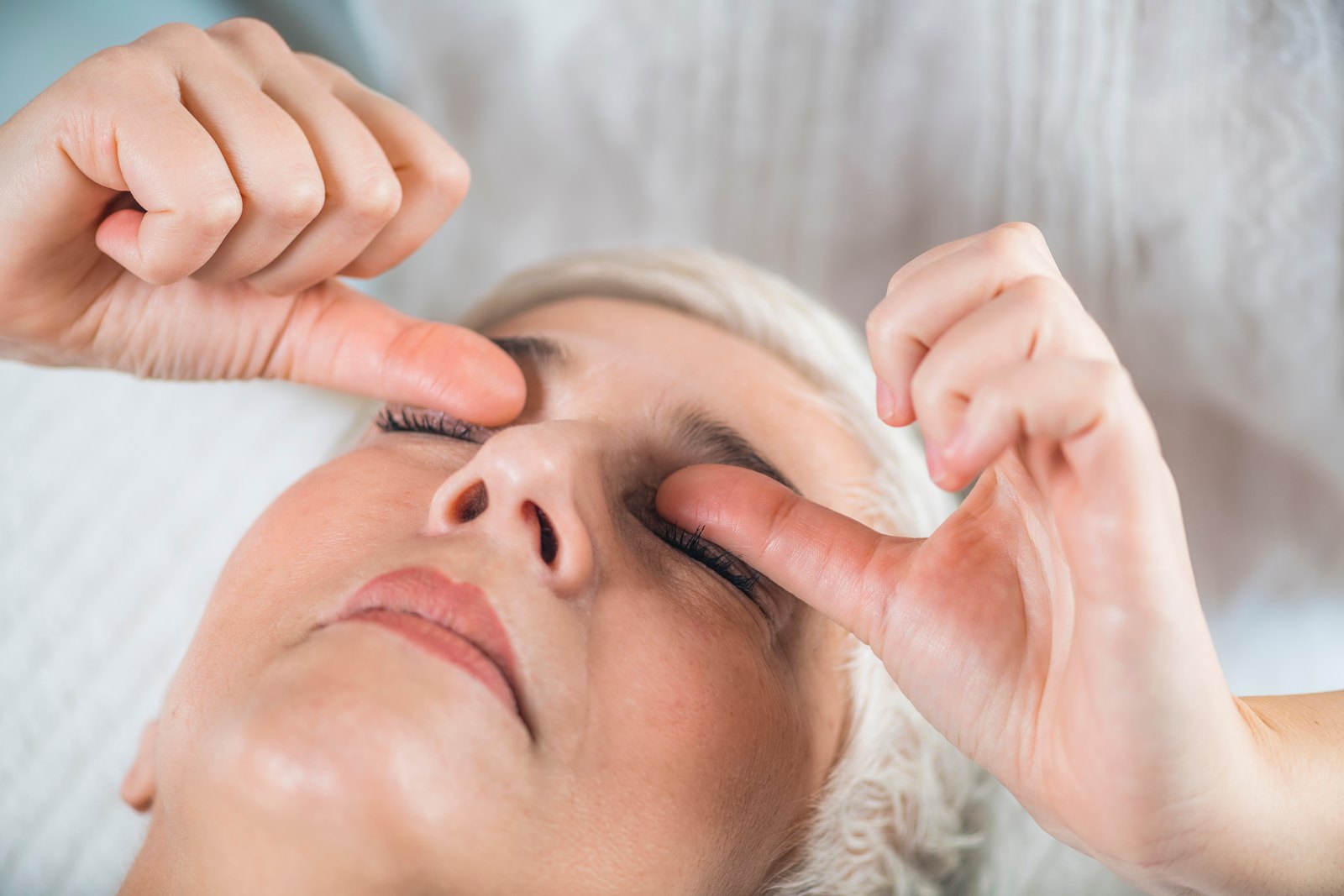
It is a comfort measure for the ocular surface and periocular tissue, performed with sterile clinic-prepared material, and nothing more. It does not reach the optic nerve and it is not a treatment for optic neuropathy. We would not offer it to anyone whose diagnosis has not been established, and never in place of urgent care.
Selected References for Scientific Support
- Jiao S, Han X. When headache meets visual disturbance: an evidence-based review of common and critical entities. Sci Prog. 2026. PubMed
- Jabri NSAA, Belushi FWMA. Delayed recognition of giant cell arteritis presenting as ear pain leading to vision loss. Oman J Ophthalmol. 2026. PubMed
- Ford JA, Gewurz D, Gewurz-Singer O. Tocilizumab in giant cell arteritis: an update for the clinician. Curr Opin Rheumatol. 2023. PubMed
- Jaen Aguila F, Gomez Escobar AJ, Navarrete Navarrete N, et al. Joint position paper on anterior ischemic optic neuropathy. Rev Clin Esp (Barc). 2026. PubMed
- Dickersin K, Li T. Surgery for nonarteritic anterior ischemic optic neuropathy. Cochrane Database Syst Rev. 2015. PubMed
- Scherer RW, Feldon SE, Levin L, et al. Visual fields at follow-up in the Ischemic Optic Neuropathy Decompression Trial. Ophthalmology. 2008. PubMed
- Foroozan R. New treatments for nonarteritic anterior ischemic optic neuropathy. Neurol Clin. 2017. PubMed
- Bennett JL, Grove NC, Johnson RK, et al. A randomized prospective trial comparing repository corticotropin injection and intravenous methylprednisolone. J Neuroophthalmol. 2023. PubMed
- Chen JJ, Flanagan EP, Pittock SJ, et al. Visual outcomes following plasma exchange for optic neuritis: an international multicenter retrospective analysis. Am J Ophthalmol. 2023. PubMed
- Liu W, Wu J, Liu Z, et al. A systematic review and meta-analysis of the efficacy and safety of eculizumab in AQP4-IgG-positive neuromyelitis optica spectrum disorder. Mult Scler Relat Disord. 2026. PubMed
- Ueda N, Kitamura Y, Baba T. A case of nutritional optic neuropathy caused by vitamin B12 and folate deficiency. Cureus. 2026. PubMed
- Nicolaou N, Nicolaou D, Douglas L, et al. Ethanol and cyanide: a case report on toxic and nutritional optic neuropathy associated with alcohol and tobacco. Cureus. 2025. PubMed
- Zainuddin MZA, Che Hamzah J, Nik Mahmood NRK, et al. Nutritional optic neuropathy: bariatric surgery gone wrong. Cureus. 2025. PubMed
- Gupta A, Tejpal T, Sriranganathan A, et al. Risk factors for ethambutol-induced optic neuropathy: a systematic review and meta-analysis of comparative studies. Can J Ophthalmol. 2026. PubMed
- Ambika S, Srilekha S. Systematic review of Leber's hereditary optic neuropathy: clinical diagnosis, genetics overview and current concepts. Indian J Ophthalmol. 2026. PubMed
- Sasagawa Y, Nakahara M, Takemoto D, et al. Optical coherence tomography detects early optic nerve damage before visual field defect in patients with pituitary tumors. Neurosurg Rev. 2023. PubMed
- Chen KY, Chan HC, Chan CM. Is acupuncture a viable therapeutic strategy for degenerative eye diseases? A systematic review and meta-analysis. Complement Ther Med. 2025. PubMed
- Saper RB, Phillips RS, Sehgal A, et al. Lead, mercury, and arsenic in US- and Indian-manufactured Ayurvedic medicines sold via the Internet. JAMA. 2008. PubMed
- Mukhopadhyay S, Abraham SE, Holla B, et al. Heavy metals in Indian traditional systems of medicine: a systematic scoping review and recommendations for integrative medicine practice. J Altern Complement Med. 2021. PubMed