Search conditions, treatments, research reviews and articles. Press Enter to see all results, Esc to close.
Search conditions, treatments, research reviews and articles. Press Enter to see all results, Esc to close.

Blog
November 11, 2026
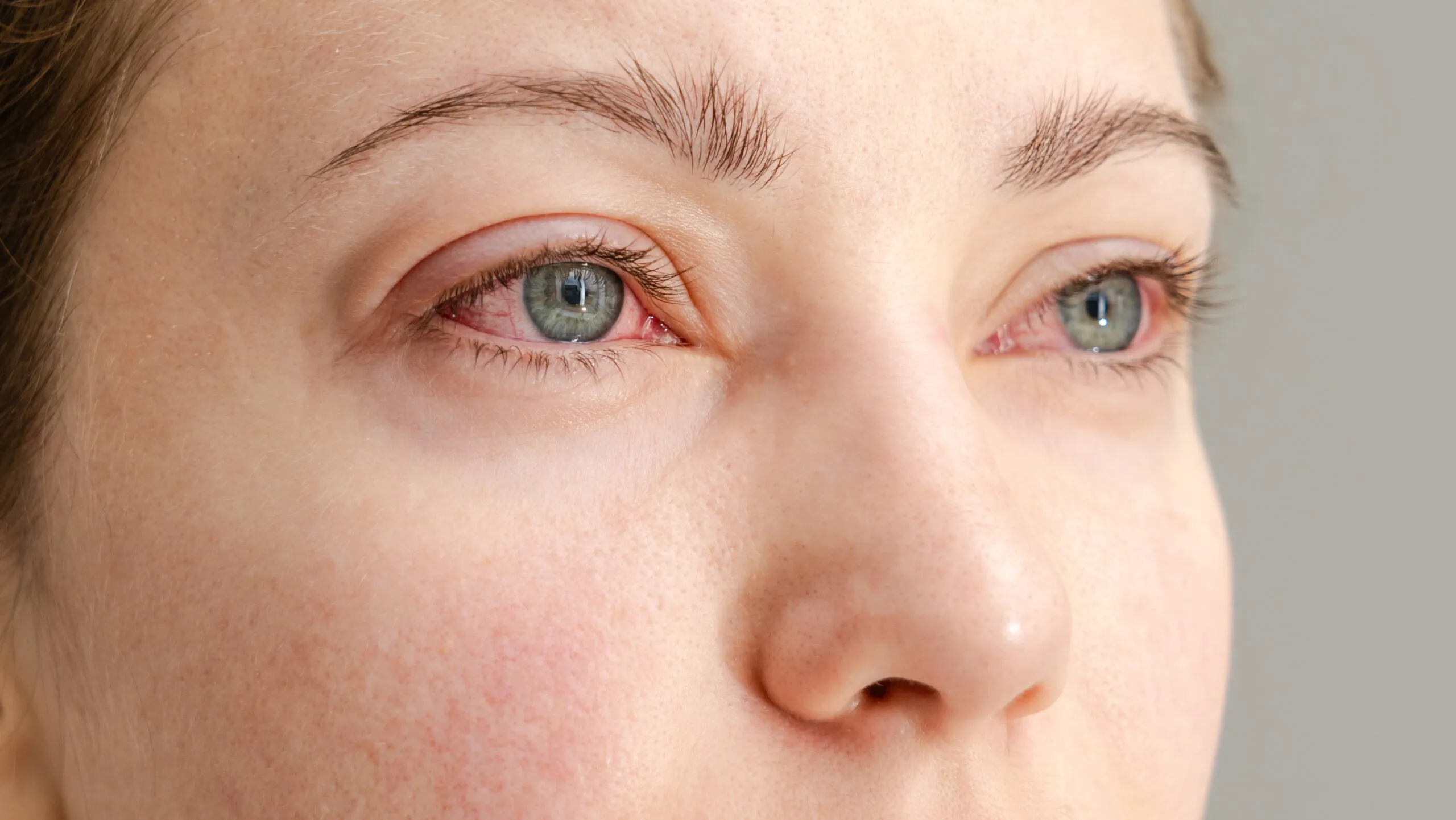
"Eye stroke" is the everyday name for vision loss caused by poor blood flow to the retina or the optic nerve. The main types are retinal artery occlusion, retinal vein occlusion and ischemic optic neuropathy, including NAION and the form caused by giant cell arteritis [1]. The usual symptom is sudden, painless loss or blurring of vision in one eye [2,7]. A blocked central retinal artery is a true stroke and a medical emergency: go to a hospital emergency department right away [2,3].
Go to an emergency department now for sudden vision loss in one eye, even if it is painless and even if it comes back after a few minutes [2,5]. Time matters: some treatments are only possible within hours, and the risk of a brain stroke is highest in the days and weeks after [2].
Key takeaways
In this article
| Type | What happens | Typical symptoms | How urgent |
|---|---|---|---|
| Central retinal artery occlusion (CRAO) | The main artery of the retina is blocked, usually by a clot or cholesterol [2] | Sudden, painless loss of vision in all of one eye [2] | Emergency, handled like a brain stroke [3] |
| Branch retinal artery occlusion (BRAO) | A smaller retinal artery is blocked [2] | Sudden loss of part of the vision in one eye; sometimes no symptoms [2] | Emergency [2] |
| Retinal vein occlusion (CRVO or BRVO) | A retinal vein is blocked, so blood and fluid leak into the retina [6] | Blurred or lost vision, sudden or over hours to days; sometimes floaters [6] | Prompt eye exam |
| NAION | Blood flow to the front of the optic nerve drops [7,8] | Painless blurring or cloudiness in one eye, often the lower part of vision, over hours to days [8] | Same day, partly to rule out giant cell arteritis [8] |
| Arteritic ischemic optic neuropathy | Inflamed arteries from giant cell arteritis cut off the optic nerve's blood supply [10] | Vision loss in someone over 50, often with headache, scalp tenderness or jaw pain when chewing [7,10] | Emergency [10] |
The most common symptom is sudden vision loss in one eye without pain [2]. Depending on the type, you may notice:
Some people notice the change on waking or only when they cover the other eye [8]. Brief vision loss in one eye that recovers within minutes, sometimes called amaurosis fugax, can be a warning of a coming stroke and also needs emergency assessment [5]. Read how this differs from ocular migraine.
Go to a hospital emergency department right away, or see an ophthalmologist immediately if one is available sooner [2]. The American Heart Association recommends that people with acute CRAO are sent straight to emergency care rather than to a routine clinic appointment [3]. Some studies link a blockage lasting just over an hour and a half to permanent vision loss [2].
For a retinal artery occlusion, doctors may try a clot-dissolving drug within several hours of the vision loss in some cases, which carries a risk of bleeding in the brain. Other approaches include breathing a carbon dioxide and oxygen mix or lowering the pressure inside the eye. None of these has been proven to work every time [2], and the AHA statement notes that some older measures, such as eye massage, may do more harm than good [3]. Because the same blockage can affect the brain, you will also have tests such as a carotid artery ultrasound, imaging of the head and neck vessels, and a heart ultrasound [2].
For a retinal vein occlusion, treatment aims at the swelling in the center of the retina. Options include anti-VEGF injections, steroid injections into the eye, and laser treatment in severe cases with abnormal new blood vessels [6]. Read more about retinal vein occlusion.
A retinal artery occlusion is usually caused by a blood clot or a piece of cholesterol that blocks the artery [2]. Risk factors are the same as for heart disease and stroke: smoking, obesity, heart or lung disease, diabetes, high cholesterol, high blood pressure, narrowing of the carotid arteries, blood clotting problems and older age. It is most common in people in their sixties [2].
Retinal vein occlusion usually affects people over 50, especially those with high blood pressure, diabetes, glaucoma or hardened arteries [6]. NAION is the most common cause of sudden optic nerve damage in people over 50 [7]. Most people with NAION have a small, crowded optic disc, and many have high blood pressure, diabetes or sleep apnea [8].
Managing these risk factors matters after any eye stroke. Care to prevent a further stroke should follow the same guidelines used after a brain stroke [3].
It depends on the type:
There is no proven treatment for NAION [8]. A large randomized trial of optic nerve sheath decompression surgery found it did not help and might be harmful, and the trial was stopped early [8]. Steroid studies have shown mixed results, with no clear difference in final vision in a double-blind trial [8]. Controlling blood pressure, diabetes and sleep apnea is widely recommended, although no randomized trial has shown that it protects the other eye [8]. The second eye is affected in about 15% to 24% of people within 5 years [8].
Because giant cell arteritis can look like NAION and can quickly blind both eyes, doctors check blood tests for inflammation in people over 50, and treat suspected arteritis with high-dose steroids straight away [8,10]. Read more about NAION.
In June 2025, the European Medicines Agency's safety committee concluded that NAION is a very rare side effect of semaglutide, the medicine in Ozempic, Rybelsus and Wegovy. It may affect up to 1 in 10,000 people taking it [9]. Large studies suggested about one extra case of NAION per 10,000 person-years of treatment in adults with type 2 diabetes [9].
If you take semaglutide and notice sudden vision loss or rapidly worsening eyesight, contact your doctor without delay. If NAION is confirmed, the medicine should be stopped [9]. Do not stop semaglutide on your own because of this risk: the American Academy of Ophthalmology and the North American Neuro-Ophthalmology Society advised patients to discuss their options with the doctor who prescribes it [8].
An eye stroke needs emergency and specialist care first. After that, Netra Eye Institute offers integrative support programs for people living with vision loss from NAION or retinal vein occlusion, alongside their ophthalmologist's care. These programs are not an emergency treatment, and they do not replace treatment from a retina specialist or neuro-ophthalmologist.
Sudden, painless loss or blurring of vision in one eye, loss of part of the visual field, blind spots or distorted lines [2]. Brief vision loss that comes back can be a warning sign too [5].
Usually not. Retinal artery occlusion and NAION are typically painless [2,8]. Pain, especially with headache, scalp tenderness or jaw pain, raises concern for giant cell arteritis [10].
Yes. A retinal artery occlusion can happen alongside a brain stroke, and the risk of a brain stroke is very high right after and for up to four weeks [2]. That is why it is treated as an emergency.
It depends on the type. Artery blockages are treated as emergencies, sometimes with clot-dissolving drugs [2]. Vein blockages are treated with injections and sometimes laser [6]. NAION has no proven treatment, so care focuses on ruling out arteritis and managing risk factors [8].
European regulators concluded that semaglutide can very rarely cause NAION, a stroke of the optic nerve, in up to 1 in 10,000 users [9]. Talk to your prescriber before making any change [8].
Sometimes. Vision after a retinal vein occlusion often improves with treatment [6]. After CRAO, fewer than 20% regain useful vision [4]. After NAION, some people improve over the first months [8].
Sources checked November 11, 2026.
This article provides general education and does not replace an individual medical or eye examination. Sudden vision loss is an emergency. Integrative care must never delay emergency or specialist treatment.