Search conditions, treatments, research reviews and articles. Press Enter to see all results, Esc to close.
Search conditions, treatments, research reviews and articles. Press Enter to see all results, Esc to close.
Blog
October 31, 2026
Corneal edema is swelling of the cornea, the clear window at the front of the eye, caused by extra fluid building up inside it. It makes vision foggy or hazy, often with glare or halos around lights, and it can hurt if small blisters form on the surface. Treatment depends on the cause: some swelling clears once the trigger is treated, while swelling from failing inner corneal cells may need a corneal transplant [1,2,3].
This article is general education, not a diagnosis. A sudden painful red eye, sudden blurry vision, or new pain or vision loss after eye surgery needs same-day or emergency care. See the warning signs below.
Key takeaways
In this article
Corneal edema is a buildup of fluid in the cornea that makes it thicker and less clear. A single layer of cells on the inner surface, the endothelium, normally pumps water out so the cornea stays thin and see-through. When those cells are damaged or overwhelmed, water collects and vision turns hazy [3,4].
The cornea has three main layers. The thin outer layer is the epithelium. The thick middle layer, the stroma, is made of evenly spaced collagen fibers, and that even spacing keeps it clear. The inner layer, the endothelium, is one cell thick [4].
When extra fluid enters the stroma, light scatters and the view looks foggy or full of glare. When fluid reaches the epithelium, small blisters called bullae can form, a stage called bullous keratopathy. The blisters can break open and cause sharp pain [3].

Get same-day or emergency care if you notice any of the following:
Do not start old steroid drops or someone else's drops. Steroids can worsen some corneal infections, such as herpes simplex infection of the surface layer [10]. For more warning signs, see our guide to urgent vision changes and eye emergency symptoms.
The main symptom is blurry, foggy or hazy vision. Many people also notice glare, halos around lights and light sensitivity. In Fuchs dystrophy, blur is often worst on waking and improves later in the day. If blisters form on the surface, the eye can feel gritty or sharply painful [1,2,3].
Why mornings? The cornea takes on a little extra water overnight, mostly in the middle layer, and thins again after you wake [11]. Weak inner cells clear that water slowly, so the haze lasts longer. At first the blur may happen only in the mornings; as the disease advances, it can last longer into the day [1,2].
Tell your eye doctor whether the main problem is blur, glare or pain, because each can point to a different problem. Glare and halos are also common with cataracts, as our article on early cataract symptoms explains.
Corneal edema happens when the inner cells cannot keep up with the water entering the cornea. Common causes are Fuchs dystrophy, eye surgery, a sudden rise in eye pressure, inflammation or infection, contact lens overwear and a failing transplant. Injuries, chemical burns and some medicines can also cause it [4,12,13].
| Cause | Typical clues | Usual next step |
|---|---|---|
| Fuchs dystrophy | Both eyes, slow change over years, blur worst on waking, symptoms usually after age 50 | Monitoring and symptom relief; transplant when vision is limited |
| After cataract or other eye surgery | Haze in the first days after surgery, or swelling that appears months later | Mild swelling often clears; lasting swelling may need a transplant |
| Acute angle-closure glaucoma | Sudden pain, red eye, halos, headache, nausea | Emergency treatment to lower eye pressure |
| Inflammation or infection (keratitis, herpes simplex) | Pain, redness, light sensitivity, often one eye | Treat the confirmed cause; steroids only under an eye doctor's care |
| Contact lens overwear | Haze or halos after long wear | Stop lens wear and get checked |
| Transplant rejection or failure | New haze, redness, light sensitivity or ache in an eye with a graft | Same-day visit; steroid drops for rejection |
| Medicines such as netarsudil (a glaucoma drop) | Honeycomb-pattern surface swelling, mostly in eyes with weak inner cells | Often clears after the eye doctor stops the drug |
These patterns are general guides. Your eye doctor confirms the cause with an exam [1,4,5,7,10,13,14].
In Fuchs dystrophy (Fuchs endothelial corneal dystrophy), the inner cells slowly stop working. It usually affects both eyes, is more common in women, and causes symptoms from around age 50 to 60. An eye doctor may see early signs years before, such as tiny bumps on the inner layer called guttae [1,2]. Netra's Fuchs dystrophy page covers its stages, tests and treatment options in more depth.
A sudden, steep rise in eye pressure can make the cornea swell. In acute angle-closure glaucoma the cornea looks hazy and vision drops, and the pressure must be lowered fast [5]. Our article on acute angle-closure glaucoma explains this emergency.
Inflammation and infection can also swell the cornea. Herpes simplex virus, for example, can inflame the inner layer and cause a round patch of swelling called disciform keratitis [10]. Treatment differs by cause, so the eye doctor needs to confirm it first. See Netra's page on keratitis (corneal inflammation).
Overwearing contact lenses can starve the cornea of oxygen and start it swelling [14]. Sleeping in lenses also raises the risk of a serious corneal infection [8]. Netarsudil, a glaucoma drop, can cause a honeycomb-pattern swelling of the surface layer, mainly in eyes with weak inner cells. It usually clears after the drug is stopped [13]. Do not stop a glaucoma drop on your own; ask the doctor who prescribed it.
Some corneal swelling is common right after cataract surgery, and mild swelling usually settles within the first few weeks as the eye heals. Swelling that does not clear, or that appears months later, usually means lasting damage to the inner cells. That type often needs a partial corneal transplant [4,14].
Cataract surgery puts stress on the endothelium. Eyes that start with fewer healthy cells, such as eyes with Fuchs dystrophy, are more likely to swell [2,4]. Other causes include a detached Descemet membrane (the thin sheet the inner cells sit on) and a rare toxic reaction to substances used in surgery [4]. A small, flat detachment often reattaches on its own within days; larger ones may be pressed back with an air or gas bubble [4].
Vision should improve step by step. Call your surgeon right away if vision gets worse after first improving, or if pain, redness or light sensitivity increases. These can be signs of infection inside the eye [6].
Before surgery, ask whether your cornea is healthy enough. One review lists two risk factors for swelling afterward: an inner-cell count below about 1,000 cells per square millimeter, and a central corneal thickness above about 640 microns [14]. Surgeons weigh these numbers with the exam and your symptoms. See when cataract surgery is needed for the wider decision.
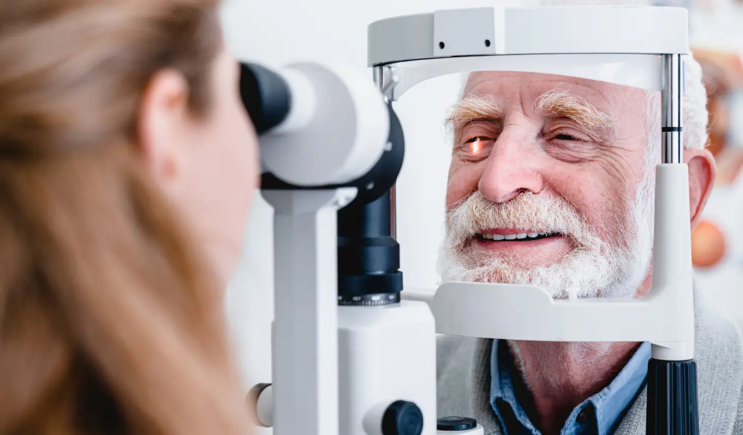
An eye doctor diagnoses corneal edema with a slit-lamp microscope, which shows haze, folds and blisters layer by layer. Pachymetry measures corneal thickness. Specular microscopy photographs the inner cells. Imaging such as anterior-segment optical coherence tomography (OCT) shows swelling and detachments in cross-section. The doctor reads these results together [2,4].
Thickness means most when it is compared with the same eye's earlier readings, because healthy corneas vary from person to person. Research often treats a rise of more than about 10% from that eye's own baseline as a sign of edema [14]. Readings taken at a similar time of day are easier to compare, because the cornea is thicker soon after waking [11].
Specular microscopy looks at only a small area, and a very cloudy cornea can make the cells hard to see. Ask your doctor which finding matters most for your plan.

Treatment targets the cause. Doctors lower high eye pressure, calm inflammation, treat infection, or stop the lens or medicine that caused the swelling. Salt (hypertonic saline) drops and ointment can ease mild swelling. When the inner cells have failed and vision stays poor, a corneal transplant that replaces the inner layer usually restores clarity [3,7,15].
Sodium chloride 5% drops, such as Muro 128, are sold over the counter for temporary relief of corneal edema. They make the tear film salty, which draws water out of the cornea [4]. The label says to use them on a doctor's advice. It also says to stop and ask a doctor if you have eye pain, vision changes, or symptoms lasting more than 72 hours [15]. They can burn briefly.
Results vary. A review found they helped most when swelling was in the middle layer, and less once blisters had formed [16]. A 2026 study in Fuchs dystrophy found extra morning drops thinned the cornea faster but did not improve vision [11]. Some doctors suggest ointment at bedtime for morning blur [4]. None of these products replace lost cells.
When surface blisters break, an eye doctor may fit a therapeutic (bandage) soft contact lens for a short time to ease pain [2,3]. Broken blisters raise the risk of infection, so these lenses need close follow-up [3]. Do not use your everyday lenses for this.
When the inner cells fail but the middle layer is still clear, a surgeon can replace just the inner layer. This is called endothelial keratoplasty. In DMEK (Descemet membrane endothelial keratoplasty), the graft is just the inner cells and their thin membrane. DSAEK (Descemet stripping automated endothelial keratoplasty) uses a slightly thicker graft [2,7].
Vision usually recovers faster after DMEK, and its rejection rate is the lowest, about 1% to 3% [7]. A Cochrane review found low-certainty evidence that DMEK gives somewhat better vision at one year. However, DMEK grafts more often need a second air bubble to reattach, called rebubbling [17]. A full-thickness transplant (penetrating keratoplasty) replaces every layer and is used when the cornea is also scarred. Full visual recovery can take up to 18 months [7].
For early Fuchs dystrophy, some surgeons remove a small central disc of the inner layer without adding donor tissue, called Descemet stripping only [2]. A review of mostly small case series found it improved vision in early disease, but about 1 in 6 eyes later needed a transplant [18].
A cloudy lens and a swollen cornea can both cause glare. Removing a cataract does not repair the cornea, and a corneal graft does not clear a cataract. Cataract surgery can make mild Fuchs dystrophy worse, so surgeons may change their technique or timing [2]. Some people have both operations at once (a triple procedure), and others have them in stages. A review found similar complication rates and vision at 6 months either way, though the evidence was low quality [19].
Call your surgeon the same day for redness, light sensitivity, eye ache or blurrier vision. These are warning signs of graft rejection, which is treated with steroid drops. A rejection episode that is severe or goes on too long can make the graft fail. While a bubble remains in the eye, follow your surgeon's travel and positioning instructions [7,20].
After DMEK or DSAEK, an air or gas bubble holds the graft in place [17]. In a lab study of donor eyes, lower air pressure, as in a plane or at altitude, sharply raised eye pressure when the bubble was large [20]. Ask your surgeon before you fly or drive over mountain passes.
A graft that does not stay attached may need rebubbling, which is a different problem from rejection or infection [7,17]. Do not start, stop or change steroid drops on your own.
Netra Restoration Therapy (NRT) is complementary care used alongside a cornea specialist, never instead of one. It has not been tested in controlled trials for corneal edema or Fuchs dystrophy. There is no evidence that it regrows inner corneal cells, clears swelling or replaces a transplant. It should never delay standard care.
Netra describes Netra Restoration Therapy as individualized care drawing on acupuncture, traditional East Asian medicine and Ayurveda. It also explains how it reviews program outcomes by diagnosis. If you are considering it for a stable, diagnosed condition, discuss timing and goals with your cornea specialist first.
Do not put oils, herbal preparations or unprescribed drops on a swollen cornea, a new graft or an eye that has just had surgery. If you choose adjunctive care, agree on one measurable goal and on which medical findings would override the plan. Morning vision changes from day to day, so one good morning cannot prove that any added treatment worked.
It depends on the cause. Mild swelling after cataract surgery, contact lens overwear, high eye pressure or inflammation often clears once the cause is treated or the eye heals. Swelling from permanently failed inner corneal cells, as in advanced Fuchs dystrophy, does not go away with drops. In that case, a corneal transplant usually restores a clear cornea.
The cornea takes on a little extra water overnight and thins again after you wake. When the inner corneal cells are weak, as in Fuchs dystrophy, that clearing takes longer, so vision can stay foggy for minutes to hours. An eye doctor can check the cornea and measure its thickness.
Usually not. Glasses correct focusing errors, but they cannot remove the light scatter caused by a swollen cornea. Swelling can also shift your prescription, so it is often better to wait until the cornea is stable before buying new glasses.
No. Corneal edema is extra fluid in the cornea, and it can clear when the cause is treated. A scar is a lasting change in the tissue after an injury or infection. Both make the cornea less clear, and an eye doctor can tell them apart with a slit-lamp exam.
Yes. Contact lenses reduce the oxygen reaching the cornea, and wearing them too long can make it swell. Sleeping in lenses also raises the risk of a serious corneal infection. If your eye becomes painful, red or blurry, take the lenses out and call your eye doctor.
No. Many people have early signs of Fuchs dystrophy for years with mild or no symptoms and only need regular checkups. A transplant is considered when swelling limits daily vision or causes painful surface blisters.
Slow, painless morning blur is usually not an emergency, but it still needs an eye exam. Sudden haze with severe eye pain, redness, halos, headache or nausea can mean acute angle-closure glaucoma and needs emergency care. New pain or vision loss after eye surgery or a corneal transplant also needs same-day care.
Sources checked October 31, 2026.
This article provides general education and does not replace an individual eye examination, diagnosis or treatment plan. If you have a sudden painful red eye with halos, headache or nausea, or new pain or vision loss after eye surgery or a corneal transplant, seek emergency or same-day care.