
Search conditions, treatments, research reviews and articles. Press Enter to see all results, Esc to close.
Search conditions, treatments, research reviews and articles. Press Enter to see all results, Esc to close.
Blog
August 9, 2026
Netra Eye Institute does not perform cataract surgery. This page is for education. If you need surgery we are glad to refer you to a cataract surgeon.
Cataracts usually do not announce themselves with a sudden white veil. Early changes appear in specific situations: headlights spread across the windshield, a restaurant menu needs a phone flashlight, colors look dull in one eye or a new prescription stops helping sooner than expected.
These experiences are valuable diagnostic data, but none belongs exclusively to cataract. The task is to connect symptoms with a visible lens opacity and rule out other causes that need different or more urgent care.
A clinician may see mild lens opacity years before it limits function. The pupil may still direct useful rays through clear lens, the fellow eye may compensate, and the visual brain adapts to gradual color or contrast change.
“Early cataract” therefore describes anatomy or functional stage, not a precise timeline. One person notices a small central posterior subcapsular opacity immediately; another tolerates broader peripheral cortical change. Symptoms depend on cataract location, pupil, task, lighting and health of cornea, retina and optic nerve.
A visible cataract does not require surgery if it is not meaningfully impairing vision or necessary examination. It does require honest monitoring and an explanation of what change should prompt review.
Normally, adding illumination improves visibility. A cataract can scatter bright light across the retinal image, creating a veil that reduces contrast. The person may read dark letters in the office but lose a pedestrian next to oncoming headlights.
Common glare situations include:
Forward scatter is not measured fully by ordinary acuity. Glare testing can sometimes demonstrate decline under a simulated source, but results vary with equipment and do not replace a task history.
Disability glare reduces the ability to detect a target because scattered light lowers retinal contrast. Discomfort glare feels unpleasant or causes squinting without necessarily reducing measured detection to the same degree. Cataracts can contribute to both, while migraine, dry eye, inflammation and neurologic sensitivity can create discomfort through other mechanisms.
Ask whether the light hurts, washes out objects, creates a halo or all three. The distinction directs examination and expectations.
A halo is a ring or glow around a light. A starburst has radiating rays. Irregular transmission through cloudy lens fibers can create both, especially around small bright sources on a dark background.
Cataract is only one cause. An unstable tear film, uncorrected astigmatism, corneal edema, keratoconus, dirty or poorly fitting contact lenses and certain IOL designs can produce similar patterns. Acute angle closure can cause halos from corneal edema together with a painful red eye, headache, nausea or vomiting—and is an emergency.
Useful details include whether halos occur in one or both eyes, improve after blinking, change with glasses or contacts, began after surgery, and accompany pain. Covering each eye separately in a safe stationary setting can localize asymmetry; do not test while driving.
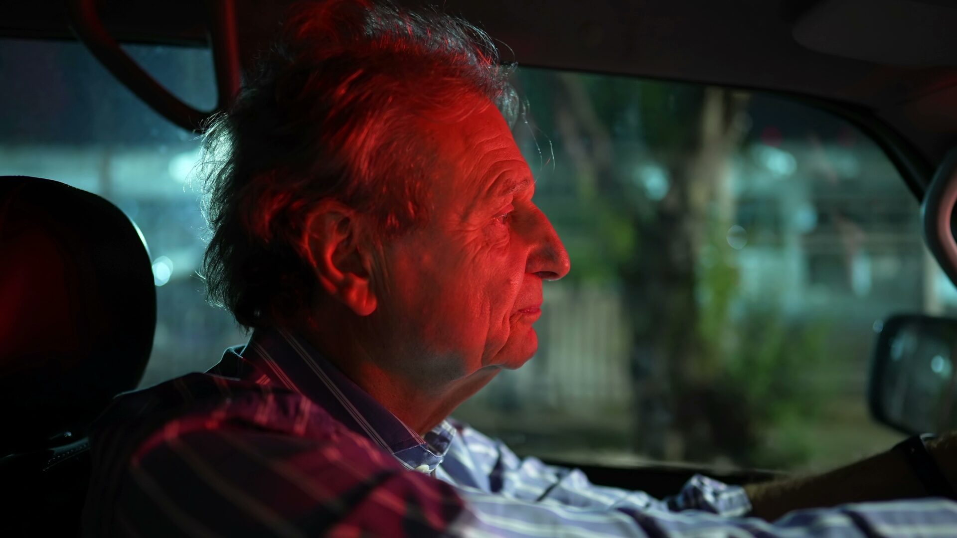
Night driving demands low-contrast detection, rapid glare recovery, peripheral awareness, motion processing and accurate judgment under changing adaptation. Cataract reduces light transmission and adds scatter, while aging pupils, retina, glaucoma, dry eye and uncorrected refractive error can also impair performance.
Warning experiences include:
These are reasons for prompt assessment and temporary driving modification. Legal acuity is a minimum, not a guarantee of safety in glare or low contrast. A clinician may discuss glare or contrast testing, field, ocular surface and driving rehabilitation depending on the problem.
Contrast sensitivity distinguishes an object from a similar background. Cataract scatter can make gray-on-gray stairs, faces in dim rooms, food on a plate or a curb in fog difficult while high-contrast letters remain readable.
Patients may call this haze, fog, washed-out vision or needing more light. The same complaint can arise from macular disease, glaucoma, corneal disease or neurologic injury. Testing and examination identify which structures contribute.
Increasing task lighting often helps until the source itself produces glare. Directed, shaded light positioned to the side is better than an exposed bulb facing the eye. Matte surfaces and contrast markings can improve safety during observation.
Progressive nuclear yellowing absorbs short-wavelength light. Blues may look greenish or gray, whites cream-colored and overall saturation reduced. Because the change is slow, the brain recalibrates and the person may not notice until comparing eyes or after first-eye surgery.
Color asymmetry can also signal optic-nerve or retinal disease, particularly when red appears washed out with pain, central blur or an afferent pupil finding. Cataract should not automatically receive credit for a disproportionate or rapid color change.
After a yellow nucleus is removed, the operated eye may see a cooler or bluish world temporarily. This commonly moderates with adaptation, but persistent new color, brightness or distortion differences deserve examination.
Nuclear sclerosis can increase lens refractive power and shift the eye toward myopia. Distance becomes blurrier while unaided near reading may briefly improve. This is not the cataract healing; it is a change in focus.
Early cataract may also increase astigmatism or create unequal refraction between eyes. Glasses can restore focus when scatter is limited, but increasingly frequent changes or a prescription that cannot reach prior clarity suggests structural limitation.
Blood-glucose fluctuation can also change refraction, sometimes over days. A person with diabetes should not assume every shift is cataract, and measurements are more reliable when glycemia is reasonably stable.
Cataract can split light into overlapping images that persist when the other eye is covered. This monocular diplopia differs from binocular double vision caused by eye misalignment, which typically resolves when either eye is covered.
Corneal irregularity, dry eye and refractive error also cause monocular ghosting. Sudden binocular diplopia, drooping lid, weakness, speech difficulty or severe headache may be neurologic and requires urgent evaluation.
A careful history saves time: Which eye? Does blinking help? Do pinhole or glasses change it? Was onset gradual? The slit lamp then tests the optical hypothesis.
As lens transmission falls, reading may require stronger illumination. Yet an unshielded bright lamp can worsen scatter. The solution is not simply higher wattage; it is controlled delivery.
Use a flexible task lamp aimed at the page from the side opposite the writing hand, shield the bulb from direct view and keep ambient room light on to reduce adaptation extremes. Increase text size and contrast before pushing brightness to discomfort.
If one eye needs dramatically more light or performance declines rapidly, schedule assessment. Retina, optic nerve and pupil function also influence luminance needs.
Entering a dim restaurant from sunlight or recovering after headlights can feel slow. Cataract reduces useful light and scatter can bleach contrast temporarily. Retinal disease, age, medication and neurologic factors also influence adaptation.
Plan extra time, use handrails and avoid stepping immediately into an unlit area. Sunglasses outdoors can reduce glare, but very dark lenses worn indoors worsen available light and adaptation. Photochromic lenses may not darken fully inside a car because the windshield blocks much ultraviolet activation.
Night blindness that is severe, lifelong, familial or disproportionate to lens opacity can signal retinal disease. It should not be dismissed as cataract.
Look for gradual distance blur, myopic prescription shift, faded or warm color and reduced dim-light contrast. Glare can occur but may be less disproportionate than with central PSC until density increases.
Symptoms often depend on pupil and ray path. Peripheral spokes can create night glare as the pupil enlarges; central extension produces broader blur and scatter. Irregular interfaces can cause ghosting.
Bright light and near work can be particularly difficult because pupillary constriction directs rays through the central posterior plaque. Glare may be severe despite modest size and progression can be faster.
Mixed cataracts combine these patterns. The clinician should not force every complaint into the dominant label when tear film or retinal disease also contributes.
Cataracts generally change gradually and painlessly. Seek urgent or same-day evaluation, depending on severity, for:
These patterns can represent angle closure, retinal tear or detachment, vascular occlusion, inflammation, infection, stroke or other emergencies. A known cataract does not protect against new disease.
Best-corrected acuity tests whether refraction improves focus. Slit-lamp examination localizes opacity. Dilation reveals peripheral lens and permits retinal and optic-nerve evaluation. Tear-film and corneal examination address fluctuating blur and halos.
Glare or brightness-acuity testing may reproduce light-dependent decline. Contrast testing can document performance beyond high-contrast letters. Macular OCT, topography, field testing or other studies are selected when symptoms or surgical planning require them.
No single test proves the cataract is the only cause. Agreement among complaint, lens location, corrected vision and the rest of the examination builds confidence that surgery will address the goal.
Before the visit, note:
Avoid repeatedly testing vision from anxiety. A concise task log over a week is more useful than dozens of unsupervised acuity checks.
Cataract is common, but corneal edema, irregular astigmatism, vitreous haze and macular dysfunction can also create fog. Haze that improves after blinking favors tear instability; haze with morning-to-evening corneal variation may suggest endothelial disease. The slit lamp localizes opacity.
Straight lines that bend or letters that warp are more characteristic of macular disease than uncomplicated lens scatter. Cataract can blur the view of a warped line, but it does not usually create a localized metamorphopsia pattern. New distortion merits dilated retinal assessment and often OCT.
A dark curtain, fixed missing patch or peripheral shadow is not an early cataract symptom. Retinal detachment, vascular disease, glaucoma or neurologic field loss must be considered. The fact that a cataract exists does not explain a new field defect.
Cataracts do not generate vitreous floaters or retinal flashes. A posterior vitreous detachment is common with age, but retinal tears must be excluded when symptoms are acute—especially with many new floaters, a curtain or vision loss.
Age-related cataracts are painless. A very advanced lens can rarely contribute to inflammation or pressure, but common early cataract should not be used to explain pain. Keratitis, uveitis, scleritis, infection and angle closure require timely differentiation.
Nuclear cataract can shift refraction, yet day-to-day or week-to-week change may reflect glucose, tear film, contact lenses or corneal disease. Stable measurements prevent unnecessary glasses remakes and improve eventual IOL calculation.
Binocular vision hides asymmetry. The clearer eye supplies detail and the brain suppresses some blur or ghosting from the worse eye. This compensation supports function, but it can delay recognition until the better eye changes.
At home, a brief safe comparison while seated can reveal brightness, color and clarity differences. Do not interpret the result as a diagnosis or perform it during driving, stairs or machinery use. A dramatic new difference deserves examination.
Clinically, acuity, refraction, pupil response and lens grade are recorded for each eye. Surgery timing and refractive target are also eye-specific. One eye may have cataract-limited vision while the other is limited by amblyopia, retina or optic nerve.
Contrast loss and glare can make stair edges, rugs and transparent doors hard to detect. Older adults may reduce activity after a near fall, leading to deconditioning and isolation. Visual symptoms should be considered alongside balance, medications, neuropathy and home hazards.
During observation, improve even illumination, add contrast to stair nosings and switches, remove loose rugs, use handrails and allow adaptation at doorways. Motion-activated night lights can help routes to the bathroom without placing an exposed source at eye level.
Cataract surgery can improve visual function and may support mobility, but fall risk is multifactorial and the interval between first- and second-eye procedures can create refractive imbalance. Plan assistance and temporary correction rather than assuming immediate binocular normalization.
Cataract does not come from computer use. Screens can reveal contrast loss, reflections and small-text difficulty, while sustained viewing worsens dry eye by reducing blink rate. Larger text, high-contrast themes, matte positioning and frequent blink breaks distinguish accommodative or surface strain from fixed scatter.
A person who can read paper but not a backlit display may be affected by brightness and glare settings. Someone who clears after a blink likely has a tear-film component. If neither adjustment nor refraction helps and a central opacity matches symptoms, cataract may be the limiting factor.
Occupational needs should be specific: color matching, microscope work, night transport, fine inspection and prolonged intermediate focus place different demands on surgery timing and IOL selection.
Brightness-acuity testing introduces a glare source while acuity is measured. Contrast-sensitivity charts use targets of decreasing contrast. Straylight instruments and questionnaires can add information in selected practices. None recreates every windshield, weather condition or neural demand.
Results can be affected by pupil, refraction, tear film, learning and device protocol. A test that shows little change does not invalidate a credible dangerous night-driving history; a poor test does not prove cataract is the sole cause.
The best decision triangulates: visible opacity in a relevant location, symptom pattern, corrected acuity or quality testing, exclusion of important comorbidity and a realistic estimate of surgical benefit.
At follow-up, “about the same” is less useful than task comparison. Can the person still drive the same route at dusk? Has font size increased? Does one eye now need repeated prescription changes? Are faces or steps harder under the same lighting?
Use consistent situations rather than risky self-testing. A short dated note can reveal progression and support shared timing. Bring current glasses and medication lists, including steroids and diabetes treatment.
Lens photographs may document structural change, but a stable photograph does not erase worsening tear film or retinal disease. Conversely, a denser lens in a comfortable non-driver may not compel immediate surgery. Anatomy and lived consequence remain partners.
If cataract truly caused glare, contrast and color change, removal often improves those domains. Outcome still depends on cornea, retina, optic nerve, IOL optics and residual refractive error. Some dysphotopsia can arise from the implant or capsule even after the original scatter is gone.
The operated eye may see colors as cooler and brighter than the unoperated eye. Dryness and mild inflammation can make early vision fluctuate. Final judgment should wait for expected healing, while increasing pain, redness or sudden decline is reported immediately.
Night-driving confidence may lag behind acuity as binocular balance and adaptation recover. Resume according to clinician advice, legal requirements and actual functional safety—not merely because surgery occurred.
Vision is a binocular perception, not two independent camera feeds displayed side by side. When one lens clouds slowly, the clearer eye contributes sharper detail and the brain weights the more useful information. Familiarity also fills gaps: a person recognizes the shape of a room or route without detecting every low-contrast feature.
This adaptation is helpful but can create a false sense that both eyes remain equal. It explains why covering the better eye during an examination can be surprising and why the untreated eye looks suddenly yellow after first-eye surgery.
Compensation has limits. It cannot reliably restore a pedestrian hidden by glare, and large inter-eye blur can reduce stereo depth or create visual discomfort. The goal of evaluation is not to defeat adaptation but to determine when it no longer supports safe, valued function.
After optical correction, the brain again adapts to brighter, clearer and sometimes differently focused input. Temporary imbalance is not unusual, particularly with monovision or a large prescription difference. Persistent difficulty needs refraction and ocular examination before being labeled a failure of neural adaptation.
Clear answers distinguish cataract treatment from treatment of everything the patient calls “vision.”
An updated refraction can improve cataract-related defocus. Anti-reflective coating reduces reflections from spectacle surfaces but cannot cancel scatter inside the lens. Clean lenses and windshield glass reduce additional stray light.
Treat ocular-surface disease with clinician-directed lubrication, lid care or prescriptions. Use larger text, higher contrast, shaded task lighting and route planning. Avoid night or rain driving when confidence or detection is impaired.
Sunglasses labeled to block UVA and UVB and a brimmed hat improve outdoor comfort and support long-term protection. Tint darkness does not determine UV blocking. Polarization reduces reflected glare but does not treat cataract.
These strategies buy functional time; they do not clear the lens. Increasing avoidance or declining safety signals that the surgery conversation should be revisited.
No oral supplement or nonapproved drop has been shown to dissolve common age-related cataract and restore transparent lens architecture. Animal, laboratory or uncontrolled findings do not establish human visual benefit.
Products marketed with N-acetylcarnosine or antioxidant claims may create expense, contamination risk or delay. Approval in another category, testimonials and a “natural” label do not prove reversal.
A balanced dietary pattern, smoking cessation, diabetes management and UV protection support health and may reduce some future risk. They are not treatments for existing symptomatic opacity. Share supplements before surgery because they can affect bleeding or anesthesia.
Surgery is considered when cataract plausibly interferes with valued activities and expected benefit exceeds individualized risk. A person does not need to fail a fixed acuity threshold or wait for a mature white lens.
Night-driving glare, work accuracy, reading endurance, falls, caregiving and inability to examine the retina are legitimate indications. The surgeon also assesses cornea, macula, optic nerve, capsule, zonules and IOL measurements.
The consultation should identify which symptom is expected to improve. If distortion comes from an epiretinal membrane or missing field from glaucoma, cataract removal may increase clarity without resolving that limitation.
Stop or restrict driving when glare recovery, sign detection or confidence is unsafe. Choose daylight, familiar routes and good weather; increase following distance; keep windows, mirrors and glasses clean; and arrange rides for night travel.
Do not compensate by staring toward oncoming lights. Direct gaze toward lane guidance while maintaining overall awareness. If central or peripheral loss remains after optical treatment, a certified driving rehabilitation specialist can assess function where available.
Family observations deserve attention, especially repeated lane errors or near misses. The aim is not to remove independence without evidence, but to prevent a crash while diagnosis and treatment proceed.
NRT at Netra Eye Institute cannot reduce lens scatter, normalize color transmission, clear a posterior plaque or replace cataract surgery. Training through degraded optical input cannot make the physical opacity transparent.
If lens opacity is mild or has been treated and a separate medically stable problem continues to affect scanning, reading, contrast or visual integration, functional assessment may determine whether NRT or another rehabilitation service is appropriate.
Any gain should be measured as task performance or strategy use, not cataract reversal. Slit-lamp examination and optical testing remain the measures of lens status.
Learn about Netra Restoration Therapy, Netra Eye Institute’s approach, cataract types and urgent vision changes.
Yes. High-contrast acuity can remain good while glare, contrast or color causes task-specific difficulty. Other causes must still be considered.
Bright point sources against darkness create scatter and demand rapid adaptation. Daytime signs provide more ambient contrast and fewer extreme luminance differences.
They reduce light reaching the eye and have not reliably proved better night-driving performance. A tint can make subjective glare feel different while reducing visibility.
Improvement after blinking suggests tear-film instability contributes. Cataract opacity does not clear with a blink, though both can coexist.
Yes, when defocus is an important component and corrected function becomes acceptable. Glasses cannot correct substantial scatter.
No. Corneal, tear-film, refractive, IOL and acute pressure problems also cause halos. Pain, redness, nausea or rapid decline is urgent.
Typical age-related cataract does not. Sudden change should be examined for retinal, corneal, pressure, vascular, inflammatory or glucose-related causes.
It cannot remove optical scatter. Environmental strategies may reduce exposure, but persistent cataract-related disability needs ophthalmic management.
Early cataract symptoms are often about quality rather than a simple acuity line: glare, halos, contrast, color, prescription stability and adaptation. Night-driving difficulty can be the most consequential expression.
Because these symptoms overlap with other disease, a comprehensive examination must show that lens opacity plausibly explains them. Optical and environmental strategies can help for a period; surgery removes the cataract when functional benefit justifies risk. NRT cannot clear the lens.
Keeping notes about the exact task, lighting and affected eye helps separate gradual lens symptoms from an urgent new retinal or neurologic change.
Medical Disclaimer: This article provides general education and is not medical advice, diagnosis, driving clearance or surgical consent. Sudden vision loss, painful redness, flashes, a curtain, marked distortion or neurologic symptoms requires urgent evaluation. Do not use supplements or unapproved drops to delay care. NRT cannot clear cataract or replace ophthalmic treatment.



