
Most clinicians who manage glaucoma have a patient they think about more than the others. The drops are in, the pressure has sat at 13 for three years, the compliance is real, and the field keeps sliding. Across the hall is the patient whose pressure was 27 on presentation and who, a decade later, has lost almost nothing. Both are open-angle glaucoma. Both are managed the same way. The disease does not seem to have read the same textbook we did.
That asymmetry is the oldest unsolved problem in glaucoma, and for most of the past seventy years the answer has been to lower pressure further and look harder. The Early Manifest Glaucoma Trial and the Collaborative Normal-Tension Glaucoma Study settled that lowering pressure slows the disease, including in eyes whose pressure was never high.1,2 What they could not settle was why pressure lowering works in some eyes and not others, or what to do for the patient in whom it plainly has not been enough.
The vascular theory of glaucoma is an attempt to answer that question. Its claim is simple: the optic nerve head is a metabolically demanding structure fed by a fragile arterial supply, and when the regulation of that supply fails, the nerve is injured whether the pressure is high or not. The theory is not new. What is new is that the perfusion deficit it describes can now be seen and measured in the clinic, and the studies of the past decade have moved it from a plausible idea to a mechanism with data behind it. This article walks through that evidence, from anatomy to imaging, and then asks what it changes about the way we examine and co-manage the patient in the chair.
Two theories, one nerve
The mechanical theory holds that raised IOP deforms the lamina cribrosa, kinks the axons passing through it, blocks axonal transport and starves the retinal ganglion cells of the neurotrophic signals that keep them alive.3 It is well supported, it explains why pressure lowering works, and it is the model most of us were trained on. The vascular theory holds that the same axons can be injured by an unstable or inadequate blood supply, and that this can happen at any pressure.4 The two are not rivals. Pressure and perfusion act on the same tissue and on each other: a higher IOP lowers ocular perfusion pressure directly, and a deformed lamina compresses the capillaries running through it. The useful way to hold both is that pressure sets how much load the nerve is under, and perfusion sets how much load it can bear. A patient who progresses at a low pressure is telling you the second number is small.
A fragile supply line
The optic nerve head is unusually exposed for a structure so important. The prelaminar and laminar tissue draws almost all its blood from the short posterior ciliary arteries, which form an incomplete ring around the nerve known as the circle of Zinn-Haller.5 The retinal, choroidal and nerve-head circulations meet here, but micro-perfusion studies show far less cross-connection between them than the classical diagrams suggest, so one territory cannot readily rescue another.6 The result is a supply with little margin. A small fall in pressure in the ciliary system, or a small rise in the resistance it has to push against, is felt at the lamina almost at once. Anatomy alone does not prove the vascular theory, but it explains why the nerve head, of all the tissues in the eye, is the one where a perfusion problem would show up first.
Dysregulation, not blockage
The instinct is to picture a vascular problem as a narrowed pipe. In glaucoma that picture is mostly wrong. Josef Flammer's 2002 synthesis, still the reference point for the field, argued that the blood flow reduction seen in glaucoma patients is rarely explained by atherosclerosis and is better explained by dysregulation: vessels that fail to hold flow steady as demand and pressure change.4 The distinction matters because a narrowed pipe delivers a constant, low flow that tissue can adapt to. A dysregulated one delivers flow that swings. Each swing below the tissue's needs is a brief ischemia; each recovery is a reperfusion, with the burst of reactive oxygen species that accompanies it. The optic nerve can survive either state for a while. It cannot survive the alternation indefinitely, and the cumulative oxidative injury to ganglion cell axons is, in this model, the lesion we call glaucoma. It also explains a clinical observation that the pipe model cannot: glaucoma patients often have normal large-vessel anatomy and abnormal peripheral microcirculation, including reduced nailfold capillary flow, which points to a systemic regulatory fault rather than a local occlusion.7
When autoregulation fails
Healthy tissue defends its blood flow. As perfusion pressure rises or falls, the arterioles constrict or dilate to keep delivery constant across a broad plateau; only at the extremes does flow begin to follow pressure. Autoregulation is what lets a healthy nerve head shrug off a hypotensive night or a high-pressure afternoon. The evidence that this defense is lost in glaucoma comes from two directions. In a primate model of chronic IOP elevation, Wang and colleagues tracked nerve-head blood flow over time and found the autoregulatory response deteriorating progressively as the disease developed, before there was major structural loss.8 In patients, Bata and colleagues used laser Doppler flowmetry while raising blood pressure with isometric exercise and found that the nerve head of a glaucomatous eye let flow rise with pressure where a healthy eye held it flat.9 The plateau, in other words, has narrowed. Once it has, every ordinary fluctuation in systemic pressure is passed straight through to the tissue. This is the mechanism that ties the systemic findings that follow, low perfusion pressure, nocturnal dips, vasospasm, to the damage at the disc.
The vasospastic patient
Some patients are built with a narrow autoregulatory plateau from the start. Flammer and Konieczka have described this as primary vascular dysregulation, or Flammer syndrome: typically a slim, often female patient with cold hands and feet, low blood pressure, a long sleep-onset time, heightened pain and smell sensitivity, and a history of migraine or Raynaud's phenomenon.10 These are not incidental findings. They are the outward signs of a vascular system that overreacts to cold, stress and emotion, and the same reactivity is present in the vessels of the nerve head. Such a patient can develop glaucomatous damage at a pressure of 14 because her nerve's tolerance for hemodynamic swings is small, and she can progress despite good pressure control because the swings continue. The syndrome is most often recognized in normal-tension glaucoma, but the trait is not confined to it. A history taken for cold extremities, migraine, low blood pressure and orthostatic symptoms costs nothing and identifies the patient in whom perfusion, not pressure, is likely to be the limiting factor.10,11
What happens overnight
The clinic sees the patient for twenty minutes in the middle of the day. The nerve head lives through the night. Two separate things happen when a patient lies down to sleep. Systemic blood pressure falls, by around 10% in most people and considerably more in "extreme dippers," a pattern that evening antihypertensive dosing can exaggerate. Independently, IOP rises in the supine position, because episcleral venous pressure rises when the head is level with the heart. Ocular perfusion pressure is approximately arterial pressure minus IOP, so at night the supply side falls while the resisting pressure rises, and perfusion pressure reaches its lowest point of the day.12 A nerve head with intact autoregulation rides this out. One without it cannot.
The epidemiology supports the physiology. A 2020 meta-analysis of population studies found that low diastolic ocular perfusion pressure, diastolic blood pressure minus IOP, was consistently associated with open-angle glaucoma.13 Nocturnal hypotension, measured by ambulatory monitoring, is among the more reproducible systemic risk factors for progression, particularly in normal-tension disease.11,12 And the newer imaging studies close the loop: Shin and colleagues showed that NTG patients with an exaggerated nocturnal diastolic dip were more likely to have focal choroidal microvasculature dropout beside the disc on OCT angiography.14 A systemic pattern the patient does not know they have leaves a visible mark on the eye. The same logic runs through a recent UK cohort that linked erectile dysfunction, an early marker of endothelial dysfunction, to a higher rate of glaucoma diagnosis (reviewed here). The practical question this raises is uncomfortable because it is so simple: for the patient progressing at target, has anyone asked when they take their blood pressure tablet?
The neurovascular unit
The most recent framing of all this drops the word "vascular" as a separate category. Neurons, astrocytes, Müller glia, pericytes and endothelial cells in the retina and nerve head form a single functional structure, the neurovascular unit, whose job is to match local blood flow to local metabolic demand moment by moment.15,16 When a ganglion cell fires, the capillaries serving it dilate within seconds; this is neurovascular coupling, and it is what the healthy retina does thousands of times a day. In glaucoma the coupling degrades. Pericytes, the contractile cells wrapped around capillaries, are lost or become dysfunctional, and the microcirculation stops answering neural demand.16 On this view, the question of whether glaucoma is a vascular disease or a neural disease is malformed: it is a disease of the partnership between the two. The framing matters for treatment because it suggests the relevant target is not blood flow in isolation, nor the neuron in isolation, but the regulatory relationship between them, which is where inflammation, oxidative stress and neurotrophic signaling all act.
Seeing perfusion
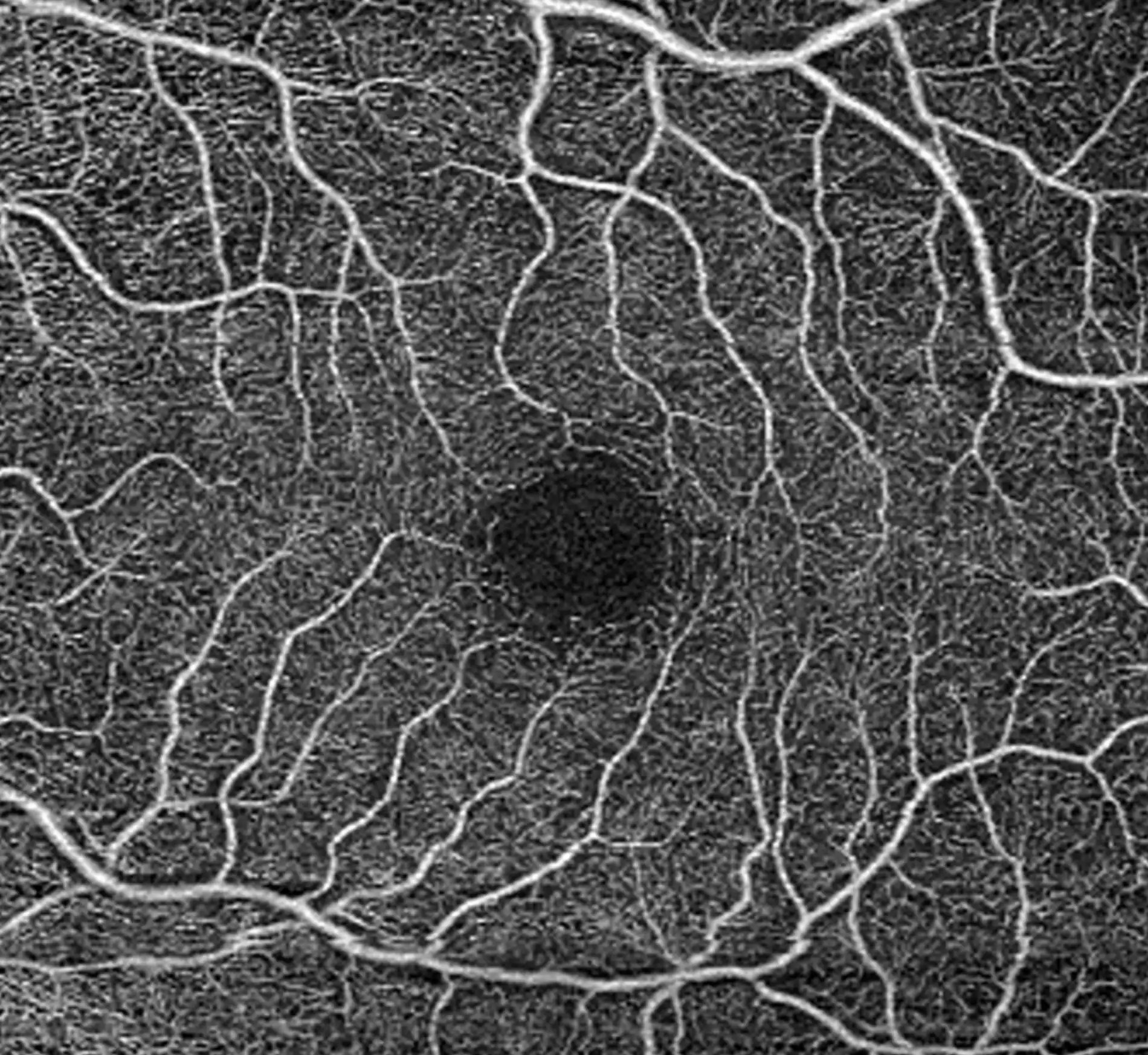
For most of its history the vascular theory suffered from a practical problem: there was no way to measure ocular perfusion in an ordinary clinic. Fluorescein angiography showed delayed filling and disc leakage in glaucomatous eyes as far back as the 1990s,17 but nobody was going to inject dye at every glaucoma visit. OCT angiography changed that. By comparing sequential B-scans and treating motion as signal, it maps flowing vessels without contrast, on the same platform that already measures the nerve fiber layer, in a few seconds per eye. The American Academy of Ophthalmology's 2021 technology assessment judged it a valid adjunct to structural OCT in glaucoma, with caveats we will come to.18
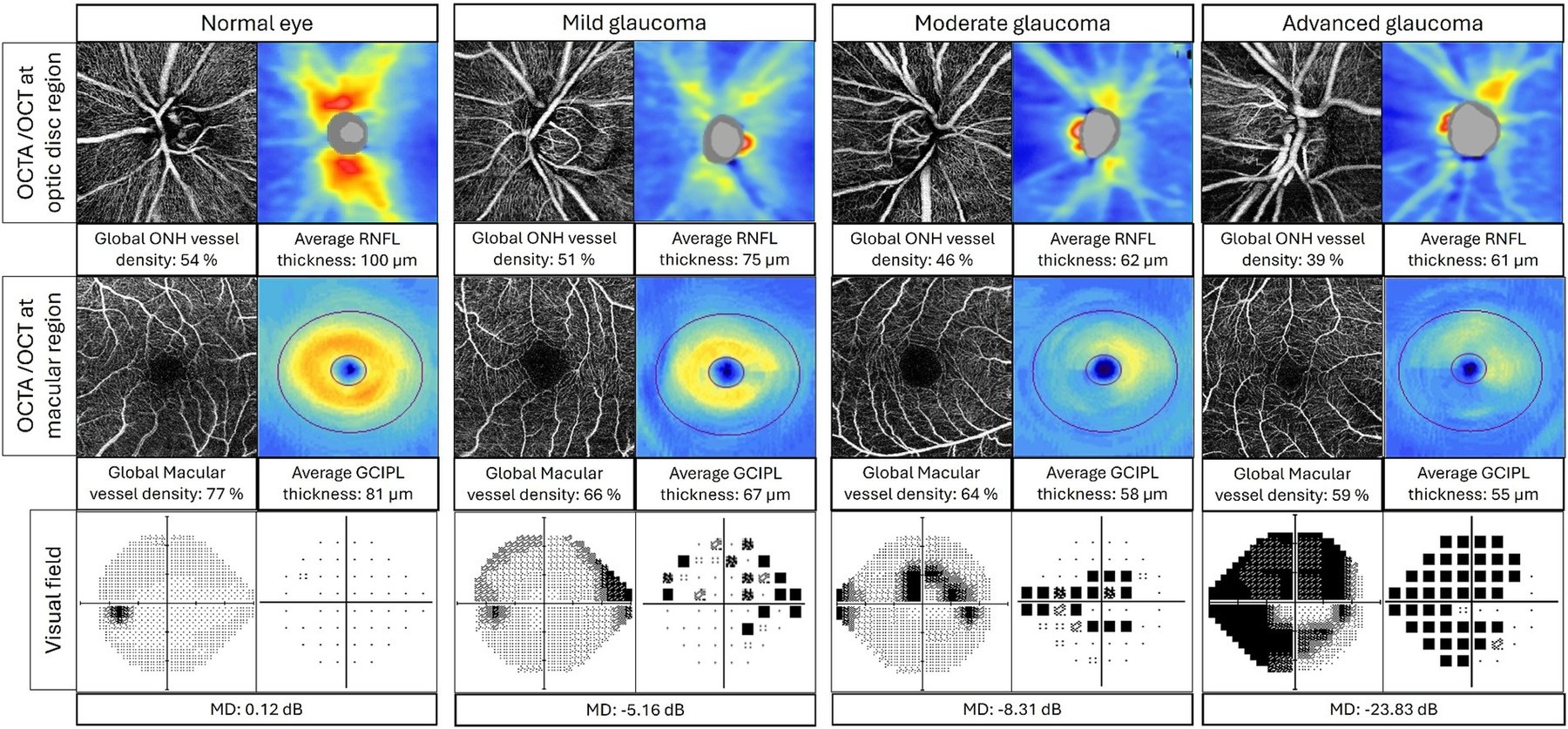
The findings are consistent. Peripapillary vessel density, the fraction of a scan area occupied by perfused vessels, falls in step with disease stage: roughly 55% in healthy eyes, 48% in mild glaucoma and 42% in moderate-to-severe disease.19 It tracks function as closely as it tracks structure; Yarmohammadi and colleagues found each 1% loss of vessel density corresponded to about 0.65 dB of visual-field mean deviation.20 It appears early. In preperimetric eyes, focal dropout of the optic disc microvasculature was present in half of Suh's series, and in most of those there was no lamina cribrosa defect to explain it, which places a vascular sign in front of the structural ones we have traditionally waited for.21,22 And it predicts. Nishida and colleagues showed that a faster initial rate of vessel density loss forecast faster subsequent field loss, and a follow-up study found that rapid early loss of nerve-head capillary density roughly doubled the risk of later progression.23,24 In advanced disease, where RNFL and ganglion cell thickness have reached their measurement floor and fields have become unreliable, vessel density keeps changing, giving the clinician one remaining metric to watch.25
Pressure lowering restores flow
If perfusion loss in glaucoma were simply the shadow of dead tissue, nothing should bring it back. The interventional studies say otherwise, and they are among the most persuasive data in this field. Liu and colleagues imaged eyes before and after glaucoma surgery that lowered IOP by a mean of about 5 mm Hg and found peripapillary capillary density recovering by roughly 12%, with the largest gains in the sectors that had the least structural damage to begin with.26 After trabeculectomy, vessel density at the level of the lamina rises as the lamina's posterior bowing relaxes, a direct demonstration that pressure was compressing the deep capillaries.27 The effect is not confined to surgery: in treatment-naive eyes started on latanoprost, the increase in peripapillary and nerve-head perfusion scaled with the size of the pressure drop, and in eyes starting above 35 mm Hg a reduction of more than half produced perfusion gains across every capillary bed measured.28,29 Two conclusions follow. There is a window, before axons are lost, in which the vascular deficit is reversible. And some of what pressure lowering achieves, it may achieve by restoring blood flow, which puts the mechanical and vascular theories on the same side of the ledger.
Normal-tension glaucoma through a vascular lens
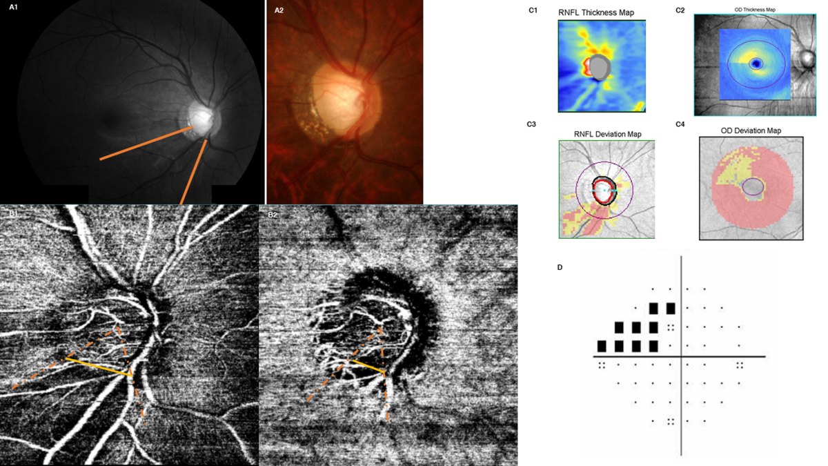
Normal-tension glaucoma is where the pressure-only model runs out of explanation, since by definition the pressure was never abnormal, and it is where the vascular findings are most striking. Perfused peripapillary capillary density is measurably lower in NTG eyes than in healthy ones, 37.8% against 43.0% in one series,30 and several groups have found that early NTG eyes show lower vessel density than POAG eyes matched for structural damage, which is hard to reconcile with the vascular loss being secondary.31,32 The most specific finding is choroidal microvasculature dropout: a complete, focal absence of choriocapillaris flow in the parapapillary region, usually inferotemporal, present in somewhere between 43% and 74% of NTG patients depending on the series.14,33 Dropout correlates with nocturnal blood pressure dipping, with worse mean deviation and with higher peak daytime IOP, which makes it read like a footprint left by the systemic problem. It is also prognostic. In a cohort of glaucoma suspects followed for conversion, baseline dropout was present in 50.7% of those who went on to develop NTG and 6.4% of those who did not, and its presence predicted faster field loss thereafter.34 It is reasonable, on this evidence, to describe NTG as a vascular disease with a mechanical component rather than the reverse, and to accept that managing it by pressure alone leaves the larger part of the problem untouched.
Reading OCT-A honestly
None of this should be taken as license to treat a vessel density number the way we treat an RNFL number. The technology is young and its failure modes are specific. Artifacts are the rule, not the exception: one systematic count found them in up to 97% of angiograms, and segmentation errors are more frequent in glaucomatous eyes than healthy ones, which is precisely where accuracy matters most.35,36 Vessel density falls almost linearly with signal strength, so a mild cataract, a dry ocular surface or an unsteady patient can produce an apparent loss of perfusion that is nothing of the kind; reliable quantification wants a signal of nine or better, which is uncommon in patients over sixty-five.37,38 Test-retest variability is roughly twice that of structural OCT, and worse in glaucomatous eyes, so a single change between visits is usually noise and confident progression needs more data points over more time.39,40 Devices differ in algorithm, scan pattern and even in what they call vessel density, so numbers do not transfer between platforms.41 Vessel density is also lowered by age, axial length, hypertension and diabetes, all of which are common in the glaucoma population, and the scan cannot tell you which is responsible.
The deepest limitation is not technical. OCT-A shows that flow is reduced; it cannot show whether reduced flow is injuring the nerve or merely reflecting the lower metabolic demand of tissue that has already been lost. The interventional and conversion data above argue for the former, but the question is not closed, and it is the right one to keep asking.
Where Netra Restoration Therapy fits
Netra Restoration Therapy (NRT) was designed around the model this article has described: an optic nerve whose survival depends on a regulated blood supply, an intact neurovascular unit and a low-inflammation, low-oxidative-stress environment, not on pressure alone. The protocol combines acupuncture, Chinese herbal medicine and Ayurvedic ophthalmic therapies chosen to act on several of those targets together, and it is given alongside, never in place of, the pressure-lowering treatment the ophthalmologist prescribes. The honest question is what the published evidence says about each component in glaucoma. The honest answer is that it points in the right direction and is preliminary throughout.
The most direct study concerns acupuncture and retrobulbar flow. Takayama and colleagues gave eleven medicated open-angle glaucoma patients a single session of body and periorbital acupuncture and measured the retrobulbar vessels with color Doppler imaging.42 The resistive index of the short posterior ciliary arteries, the very vessels that supply the circle of Zinn-Haller, fell significantly against a control day, as did IOP, with no change in systemic blood pressure or heart rate. It is a case series of one session, and the authors say so. The 2020 Cochrane review of acupuncture for glaucoma concluded that the evidence is insufficient to determine benefit, which is a statement about trial size and design, not a finding of no effect.43
The herbal evidence is mixed and worth stating plainly. Ginkgo biloba extract, a standard vasoactive botanical in both TCM-informed and Western integrative practice, was associated with a slower rate of visual-field decline in a retrospective Korean series of 42 NTG patients followed for six years, from 0.62 to 0.38 dB per year,44 but a four-week randomized crossover trial in 28 Chinese NTG patients found no effect on mean deviation or contrast sensitivity.45 Four weeks cannot detect a change in progression rate, and a retrospective series cannot exclude regression to the mean; together the two studies suggest an effect that is modest and slow if it exists at all.
The strongest randomized evidence for a non-drug intervention bears directly on vascular dysregulation. Dada and colleagues randomized 90 medicated POAG patients to three weeks of daily mindfulness meditation or medication alone.46 IOP in the meditation group fell by about 6 mm Hg, roughly a third, and serum cortisol, IL-6, TNF-α and reactive oxygen species fell while BDNF rose, a biomarker pattern that maps closely onto the inflammatory and neurotrophic targets NRT is built around. Netra's glaucoma protocol includes a structured relaxation and breathing component for this reason. Flammer's own group, notably, recommends the same orientation for the vasospastic patient: avoid hypotension, review evening antihypertensives, correct magnesium deficiency, and reach for gentle systemic measures before pharmacological ones.10
What none of this amounts to is a trial of NRT itself, and Netra does not claim one. The mechanisms NRT targets are the mechanisms this article has described; its components each have small studies pointing the right way; and the outcome data needed to move from plausible to proven have not yet been collected. That is why Netra publishes its rationale openly and invites referring clinicians to collaborate on outcome research. In the meantime, the therapy's proper place is the one the vascular theory itself suggests: adjunctive care for a patient whose pressure is treated and whose perfusion is still in question.
Takeaways for the clinic
The optic nerve head is fed by a supply line with little redundancy, defended by an autoregulatory system that glaucoma erodes, and exposed every night to the lowest perfusion pressure of the day. None of that shows up on a tonometer. What follows is not a new drug but a wider history and a sharper eye.
- Ask the vascular questions. In any patient progressing at target, and in every NTG patient: cold hands and feet, migraine, Raynaud's, low or labile blood pressure, orthostatic symptoms, sleep apnea, and when they take their blood pressure medication.
- Do the subtraction. Diastolic blood pressure minus IOP takes seconds and identifies the patient with a low perfusion margin.
- Think about the night. Where nocturnal hypotension is plausible, ambulatory blood pressure monitoring and a note to the primary care physician about moving evening doses to the morning is a low-risk, well-reasoned step.
- Use OCT-A for what it is good at. Risk stratification in preperimetric eyes, a signal in advanced disease past the structural floor, and a reason to escalate or hold. Read it with its artifact rate and signal dependence in mind, and compare only against the same device over time.
- Keep lowering pressure. It remains the only intervention with trial evidence, and part of what it does is restore perfusion.
- Treat the eye as part of a circulatory system. Co-manage the systemic side with the same seriousness as the pressure, and consider adjunctive approaches that target regulation, inflammation and neurotrophic support, with clear eyes about how much evidence each one has.
For seventy years glaucoma has been managed by the number we could measure. We can now measure the other one. The work ahead is learning what to do with it.
References
- Heijl A, Leske MC, Bengtsson B, et al. Reduction of intraocular pressure and glaucoma progression: results from the Early Manifest Glaucoma Trial. Arch Ophthalmol. 2002;120(10):1268-79.
- Collaborative Normal-Tension Glaucoma Study Group. Comparison of glaucomatous progression between untreated patients with normal-tension glaucoma and patients with therapeutically reduced intraocular pressures. Am J Ophthalmol. 1998;126(4):487-97.
- Ahmad SS. The mechanical theory of glaucoma in terms of prelaminar, laminar, and postlaminar factors. Taiwan J Ophthalmol. 2023;14(3):376-86.
- Flammer J, Orgül S, Costa VP, et al. The impact of ocular blood flow in glaucoma. Prog Retin Eye Res. 2002;21(4):359-93.
- Onda E, Cioffi GA, Bacon DR, Van Buskirk EM. Microvasculature of the human optic nerve. Am J Ophthalmol. 1995;120(1):92-102.
- Yu PK, McAllister IL, Morgan WH, et al. Inter-relationship of arterial supply to human retina, choroid and optic nerve head using micro perfusion and labeling. Invest Ophthalmol Vis Sci. 2017;58(10):4309-19.
- Cousins CC, Chou JC, Greenstein SH, et al. Resting nailfold capillary blood flow in primary open-angle glaucoma. Br J Ophthalmol. 2019;103(2):203-7.
- Wang L, Cull G, Burgoyne CF, et al. Longitudinal alterations in the dynamic autoregulation of optic nerve head blood flow revealed in experimental glaucoma. Invest Ophthalmol Vis Sci. 2014;55(8):5117-26.
- Bata AM, Fondi K, Witkowska KJ, et al. Optic nerve head blood flow regulation during changes in arterial blood pressure in patients with primary open-angle glaucoma. Acta Ophthalmol. 2019;97(7):e1000-e1005.
- Konieczka K, Flammer J. Treatment of glaucoma patients with Flammer syndrome. J Clin Med. 2021;10(15):3388.
- Choi J, Kook MS. Systemic and ocular hemodynamic risk factors in glaucoma. Biomed Res Int. 2015;2015:141905.
- Chung HJ, Hwang HB, Lee NY. The association between primary open-angle glaucoma and blood pressure: two aspects of hypertension and hypotension. Biomed Res Int. 2015;2015:827516.
- Kim KE, Oh S, Baek SU, et al. Ocular perfusion pressure and the risk of open-angle glaucoma: systematic review and meta-analysis. Sci Rep. 2020;10(1):10056.
- Shin JW, Jo YH, Song MK, Won HJ, Kook MS. Nocturnal blood pressure dip and parapapillary choroidal microvasculature dropout in normal-tension glaucoma. Sci Rep. 2021;11(1):206.
- Wareham LK, Calkins DJ. The neurovascular unit in glaucomatous neurodegeneration. Front Cell Dev Biol. 2020;8:452.
- Alarcon-Martinez L, Shiga Y, Villafranca-Baughman D, et al. Neurovascular dysfunction in glaucoma. Prog Retin Eye Res. 2023;97:101211.
- Arend O, Remky A, Plange N, et al. Fluorescein leakage of the optic disc in glaucomatous optic neuropathy. Graefes Arch Clin Exp Ophthalmol. 2005;243(7):659-64.
- WuDunn D, Takusagawa HL, Sit AJ, et al. OCT angiography for the diagnosis of glaucoma: a report by the American Academy of Ophthalmology. Ophthalmology. 2021;128(12):1775-85.
- Lee EJ, Lee KM, Lee SH, et al. OCT angiography of the peripapillary retina in primary open-angle glaucoma. Invest Ophthalmol Vis Sci. 2016;57(14):6265-70.
- Yarmohammadi A, Zangwill LM, Diniz-Filho A, et al. Relationship between optical coherence tomography angiography vessel density and severity of visual field loss in glaucoma. Ophthalmology. 2016;123(12):2498-508.
- Suh MH, Weinreb RN, Zangwill LM. Optic disc microvasculature dropout in preperimetric glaucoma. J Glaucoma. 2024;33(7):490-8.
- Suh MH, Jung DH, Weinreb RN, et al. Optic disc microvasculature dropout in glaucoma detected by swept-source optical coherence tomography angiography. Am J Ophthalmol. 2022;236:261-70.
- Nishida T, Moghimi S, Wu JH, et al. Association of initial optical coherence tomography angiography vessel density loss with faster visual field loss in glaucoma. JAMA Ophthalmol. 2022;140(2):160-8.
- Tansuebchueasai N, Nishida T, Moghimi S, et al. Rate of initial optic nerve head capillary density loss and risk of visual field progression. JAMA Ophthalmol. 2024;142(3):210-18.
- Moghimi S, Bowd C, Zangwill LM, et al. Measurement floors and dynamic ranges of OCT and OCT angiography in glaucoma. Ophthalmology. 2019;126(7):980-8.
- Liu L, Takusagawa HL, Greenwald MF, et al. Optical coherence tomographic angiography study of perfusion recovery after surgical lowering of intraocular pressure. Sci Rep. 2021;11(1):17251.
- Kim JA, Kim TW, Lee EJ, et al. Microvascular changes in peripapillary and optic nerve head tissues after trabeculectomy in primary open-angle glaucoma. Invest Ophthalmol Vis Sci. 2018;59(11):4614-21.
- Liu C, Umapathi RM, Atalay E, et al. The effect of medical lowering of intraocular pressure on peripapillary and macular blood flow as measured by optical coherence tomography angiography in treatment-naive eyes. J Glaucoma. 2021;30(6):e291-e298.
- Holló G. Influence of large intraocular pressure reduction on peripapillary OCT vessel density in ocular hypertensive and glaucoma eyes. J Glaucoma. 2017;26(3):e7-e10.
- Scripsema NK, Garcia PM, Bavier RD, et al. Optical coherence tomography angiography analysis of perfused peripapillary capillaries in primary open-angle glaucoma and normal-tension glaucoma. Invest Ophthalmol Vis Sci. 2016;57(9):OCT611-20.
- Lee MW, Yu HY, Park KS, et al. A comparison of peripapillary vessel density between subjects with normal-tension glaucoma and primary open-angle glaucoma with similar extents of glaucomatous damage. Sci Rep. 2023;13(1):4567.
- Zehavi-Dorin T, Kutzscher AE, Badr M, et al. A comparison of vessel density in primary open-angle glaucoma and normal-tension glaucoma patients using OCT angiography. Clin Ophthalmol. 2025;19:101-10.
- Shin JW, Lee JY, Lee BJ, et al. Clinical characteristics of choroidal microvasculature dropout in normal-tension glaucoma versus nonarteritic anterior ischemic optic neuropathy: an optical coherence tomography angiography study. Sci Rep. 2021;11(1):5120.
- Jung Y, Park HL, Shin H, et al. Microvasculature dropout and development of normal tension glaucoma in glaucoma suspects: the normal tension glaucoma suspect cohort study. Am J Ophthalmol. 2022;236:112-21.
- Holmen IC, Konda SM, Pak JW, et al. Prevalence and severity of artifacts in optical coherence tomographic angiograms. JAMA Ophthalmol. 2020;138(2):119-26.
- Enders C, Lang G, Dreyhaupt J, et al. Quantity and quality of image artifacts in optical coherence tomography angiography. PLoS One. 2019;14(1):e0210505.
- Yu JJ, Camino A, Liu L, et al. Signal strength reduction effects in OCT angiography. Ophthalmol Retina. 2019;3(10):835-42.
- Lim HB, Kim YW, Kim JM, et al. The importance of signal strength in quantitative assessment of retinal vessel density using optical coherence tomography angiography. Sci Rep. 2018;8(1):12897.
- Nishida T, Moghimi S, Hou H, et al. Long-term reproducibility of optical coherence tomography angiography in healthy and stable glaucomatous eyes. Br J Ophthalmol. 2023;107(5):657-62.
- Lee JC, Grisafe DJ, Burkemper B, et al. Intrasession repeatability and intersession reproducibility of peripapillary OCTA vessel parameters in non-glaucomatous and glaucomatous eyes. Br J Ophthalmol. 2021;105(11):1534-41.
- Rao HL, Pradhan ZS, Suh MH, et al. Optical coherence tomography angiography in glaucoma. J Glaucoma. 2020;29(4):312-21.
- Takayama S, Seki T, Nakazawa T, et al. Short-term effects of acupuncture on open-angle glaucoma in retrobulbar circulation: additional therapy to standard medication. Evid Based Complement Alternat Med. 2011;2011:157090.
- Law SK, Wang L, Li T. Acupuncture for glaucoma. Cochrane Database Syst Rev. 2020;2(2):CD006030.
- Lee J, Sohn SW, Kee C. Effect of Ginkgo biloba extract on visual field progression in normal tension glaucoma. J Glaucoma. 2013;22(9):780-4.
- Guo X, Kong X, Huang R, et al. Effect of Ginkgo biloba on visual field and contrast sensitivity in Chinese patients with normal tension glaucoma: a randomized, crossover clinical trial. Invest Ophthalmol Vis Sci. 2014;55(1):110-6.
- Dada T, Mittal D, Mohanty K, et al. Mindfulness meditation reduces intraocular pressure, lowers stress biomarkers and modulates gene expression in glaucoma: a randomized controlled trial. J Glaucoma. 2018;27(12):1061-7.
Source
Netra Eye Institute. Looking Beyond IOP in Glaucoma: The Vascular Side of the Puzzle. Netra Clinical Knowledge Hub, Research Reviews. September 2026. 46 references; full list at the end of the article.
Read the journal articleThis article was prepared by the Netra Eye Institute editorial team together with clinicians in the relevant specialty. AI tools may have been used during drafting and editing; every statement was reviewed and approved by the human editors responsible for this series.