

Glaucoma damages the optic nerve, and that damage does not come back. Lowering intraocular pressure is the only intervention shown in randomized trials to slow it, and no Ayurvedic therapy has been shown to lower intraocular pressure. If you have glaucoma, drops, laser, or surgery are the treatment. Everything else sits around that treatment and supports it. What integrative care can honestly offer here is narrower and more specific than most clinics admit: stress physiology, sleep and vascular health, ocular surface tolerance of long-term drops, and the unglamorous work of staying on therapy for decades.
Complementary-care boundary: Ayurvedic eye care does not replace prostaglandin analogs, beta-blockers, alpha agonists, carbonic anhydrase inhibitors, or rho kinase inhibitors. It does not replace selective laser trabeculoplasty, minimally invasive glaucoma surgery, trabeculectomy, tube shunt implantation, or laser peripheral iridotomy for a narrow angle. It does not replace tonometry, gonioscopy, optic nerve imaging, or visual field testing on the schedule your ophthalmologist sets. Nothing offered at Netra Eye Institute is a substitute for any of these, and delaying them costs vision that cannot be recovered.
An Integrative Approach to the Drivers of Glaucoma
Glaucoma is a progressive optic neuropathy. Retinal ganglion cells and their axons die, the optic disc excavates, and the visual field thins from the periphery inward. Intraocular pressure is the dominant risk factor and the only one we can treat, but it is not the whole story. Many patients progress at statistically normal pressures, and many people with high pressures never develop damage. That gap is where blood flow, nocturnal blood pressure, corneal biomechanics, and individual susceptibility of the optic nerve head come in.
An integrative approach does not mean adding herbs to a pressure problem. It means taking the parts of a patient's life that genuinely interact with the disease and managing them properly: untreated sleep apnea, aggressive nighttime antihypertensive dosing, chronic sympathetic arousal, an ocular surface so inflamed that drops become intolerable, and the practical realities that cause people to skip doses. These are real clinical levers. None of them substitutes for pressure control.
Netra Eye Institute works as a supportive service alongside your glaucoma specialist, not in place of one. We do not set target pressures, we do not adjust your medications, and we do not monitor your disease. Your ophthalmologist does that. Our role is to make the rest of your care easier to sustain and to be candid with you about what Ayurvedic therapy can and cannot do in a disease this serious.
Why Glaucoma Care Should Be Multi-Factorial
Pressure is the only driver with proven treatment benefit
This is the fixed point around which everything else moves. Randomized trials, including the Ocular Hypertension Treatment Study, the United Kingdom Glaucoma Treatment Study, and the LiGHT trial, all show the same thing: lowering intraocular pressure reduces the rate of conversion and progression. No other intervention has that record. Any care plan that treats pressure as optional is not a glaucoma plan.
Damage happens at pressures that look normal
A sizeable share of open-angle glaucoma occurs with pressures that never exceed 21 mmHg. That does not make pressure irrelevant; in normal-tension glaucoma, lowering pressure further still slows progression. It does mean a single normal reading is not reassurance, and it explains why some patients need target pressures in the low teens while others do well at 18.
Perfusion of the optic nerve head matters
The optic nerve head sits at the intersection of intraocular pressure and blood pressure. When nighttime blood pressure dips sharply, perfusion pressure at the disc falls, and some patients progress despite good tonometry. Vascular dysregulation, cold extremities, and migraine cluster with this pattern. These findings are associations rather than proven treatment targets, but they shape how we review a patient's systemic medications.
Structure and function do not fail in step
Optic nerve fiber layer thinning on OCT and visual field loss on perimetry track each other loosely. Early disease often shows structural change first; advanced disease shows functional change first, because the OCT reaches a measurement floor. Following both, over years, is how progression is actually detected. One test alone will mislead you in one direction or the other.
The disease is silent until it is far along
Peripheral loss does not feel like a curtain or a blur. The brain fills in missing field, and most patients notice nothing until a large fraction of the nerve is gone. This is why screening, family history, and keeping appointments matter more in glaucoma than in almost any other eye condition. Symptoms are a late and unreliable guide.
Adherence is the quiet failure point
A drop that is not instilled has no effect. Real-world adherence to glaucoma medication is poor, and the reasons are mundane: stinging, redness, cost, complicated schedules, arthritis, forgetting, and simply not feeling sick. A meaningful part of what supportive care contributes in glaucoma is making the prescribed regimen tolerable and habitual. That is not glamorous, and it is probably the highest-value thing we do.
Key Biological Mechanisms in Glaucoma
Resistance in the conventional outflow pathway
Aqueous humor is produced by the ciliary body and drains mainly through the trabecular meshwork into Schlemm canal. In open-angle glaucoma the meshwork stiffens, its cells are lost, and extracellular matrix accumulates, raising outflow resistance. Pressure rises because drainage falls, not because production surges. Laser trabeculoplasty and rho kinase inhibitors both act on this tissue.
Mechanical strain at the lamina cribrosa
Axons leaving the eye pass through a perforated collagen sieve called the lamina cribrosa. Pressure differences across this structure deform it, and the deformation is not uniform. Where strain concentrates, axonal bundles are pinched. This is the leading explanation for why glaucomatous field loss follows arcuate patterns that match nerve fiber anatomy rather than a random scatter of dots.
Failure of axonal transport
Retinal ganglion cells depend on transport of mitochondria and neurotrophic factors along axons that run centimeters to the brain. Strain at the lamina interrupts that traffic in both directions. Deprived of survival signaling from their targets, cells begin a programmed death sequence. Early in this process the cell is stressed but alive, which is why timely pressure reduction can preserve function that is not yet lost.
Glial remodeling and neuroinflammation
Astrocytes and microglia in the optic nerve head respond to mechanical and metabolic stress by changing shape, migrating, and releasing inflammatory mediators. Some of this response is protective and some accelerates tissue remodeling. This is an active research area, and it is the honest basis for describing glaucoma as a neurodegenerative process rather than a plumbing problem alone.
Oxidative stress and mitochondrial load
Ganglion cell axons are unmyelinated within the retina and carry a heavy mitochondrial burden per unit length. That makes them metabolically expensive and vulnerable to oxidative injury. Trabecular meshwork cells show oxidative damage as well. Antioxidant supplementation is often proposed on this reasoning, but a systematic review of nutritional supplementation in glaucoma found the human evidence thin and inconsistent.
Autonomic tone, cortisol, and pressure
Sympathetic arousal and glucocorticoid signaling both influence aqueous dynamics, and elevated cortisol is associated with higher intraocular pressure. This is the one mechanism in this list with a plausible line running from a behavioral intervention to a measurable pressure change, which is why the mindfulness trials discussed later are worth taking seriously rather than dismissing as relaxation advice.

How Glaucoma Presents and How It Progresses
Primary open-angle glaucoma
The common form, and the quiet one. The drainage angle is anatomically open, pressure rises gradually, and the nerve thins over years. There is no pain, no redness, and usually no symptom until mid-peripheral field is substantially lost. Most patients are found on a routine exam or because a relative was diagnosed. Progression rates vary from negligible to rapid.
Angle-closure and the acute attack
When the iris blocks the drainage angle, pressure can climb into the 40s or higher within hours. The presentation is unmistakable: severe eye pain, a red eye, blurred vision with halos around lights, headache, and often nausea and vomiting. This is an emergency. Untreated, it can destroy vision in a day. It is treated with pressure-lowering medication and laser peripheral iridotomy, sometimes lens extraction.
Normal-tension glaucoma
Characteristic optic nerve and field damage with pressures consistently in the statistically normal range. Vascular factors, nocturnal hypotension, and sleep apnea appear more often in this group. Treatment is still pressure reduction, aiming for a substantially lower target than the starting point, because that remains the only approach with trial support.
Secondary glaucomas
Pressure rises because of another process. Pseudoexfoliation deposits clog the meshwork and tend to produce higher, more unstable pressures. Pigment dispersion does something similar in younger myopic patients. Steroid use, uveitis, trauma, and neovascularization from diabetic or vascular retinal disease all cause secondary glaucoma. The underlying cause has to be treated alongside the pressure.
Rate of progression varies enormously
Two patients with the same visual field at diagnosis can have very different futures. Some lose a fraction of a decibel per year and never become visually impaired; others lose several times that and need aggressive surgery. This is why serial testing over time, not a single snapshot, drives treatment decisions, and why the interval between visits is a clinical judgment rather than a formality.
What advanced disease looks like
Late glaucoma is disabling in a specific way. Central acuity can stay strong while the field narrows to a tunnel, so patients read the bottom line of the chart and still bump into doorframes, miss curbs, and lose the ability to drive safely. Contrast sensitivity and dark adaptation degrade. Preventing this is the entire point of treatment, because none of it is recoverable.
How Glaucoma Is Assessed at Netra Eye Institute
We do not duplicate your glaucoma specialist's monitoring, and we do not replace it. What we do is read the data you already have, look for the supportive factors that often go unexamined, and make sure nothing in a complementary plan interferes with your treatment. If you do not yet have a glaucoma specialist, arranging that comes before anything else we might offer.
Intraocular pressure and its variation
A single reading is one point on a curve that moves through the day, often peaking in the early morning. We review your recorded pressures over time rather than one number, and note whether readings were taken at different hours. Where progression is occurring despite apparently controlled pressure, diurnal measurement by your specialist is a reasonable next step.
Corneal thickness and biomechanics
Goldmann tonometry assumes an average cornea. A thin cornea causes pressure to read lower than it truly is, and a thick one the reverse. Central corneal thickness was one of the strongest predictors of conversion in the Ocular Hypertension Treatment Study. We confirm this measurement exists in your record, because without it your readings cannot be interpreted properly.
Gonioscopy and angle status
Whether your drainage angle is open, narrow, or closed changes everything about risk and about which therapies are safe. Angle status also determines whether certain dilating drops and some systemic medications are hazardous for you. We check that gonioscopy has been performed and recorded, and we flag it when it has not.
Optic nerve head and nerve fiber imaging
OCT quantifies retinal nerve fiber layer and ganglion cell complex thickness and compares it with normative data and with your own prior scans. Disc photographs remain useful for documenting hemorrhages and rim contour. We review trend reports rather than single scans, since measurement noise easily masquerades as change.
Visual field testing and progression analysis
Standard automated perimetry is the functional benchmark. Early fields are unreliable while a patient learns the test, so we look at the whole series and at the guided progression analysis rather than reacting to one poor result. Fields that keep worsening despite stable pressures are a reason to return to your specialist, not a reason to try something alternative.
Medication review and drop technique
We ask you to demonstrate how you instill your drops. Missing the eye, contaminating the bottle tip, and instilling two drops of the same medication are all common and all consequential. We also check for interactions, including systemic beta-blocker use and any medication with anticholinergic effects in a patient with narrow angles.
Systemic, vascular, and sleep review
We ask about blood pressure control and the timing of antihypertensive doses, about snoring and daytime sleepiness, about migraine and cold hands, and about steroid exposure in any form, including inhalers, skin creams, and joint injections. Obstructive sleep apnea is associated with glaucoma in meta-analysis and is worth diagnosing on its own merits.
Ocular surface assessment
Years of preserved drops damage the ocular surface, and a patient with burning, gritty eyes is a patient who will start skipping doses. We assess tear film stability, meibomian gland function, and surface staining, because fixing this is one of the few places where supportive care demonstrably improves glaucoma outcomes, by protecting adherence.
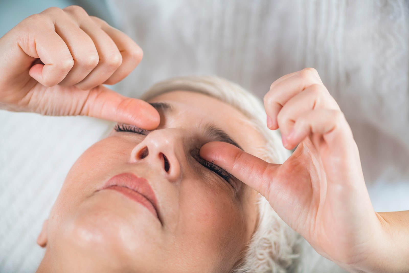
What Ayurvedic Eye Care Offers in Glaucoma
The honest summary is that Ayurveda has no trial evidence for glaucoma. There is no randomized controlled trial showing that any classical Ayurvedic formulation, oil therapy, or purification protocol lowers intraocular pressure or slows optic nerve damage. Searches of the published literature return case reports and mechanistic speculation, not outcome trials. We will not present that absence as an open question in your favor.
What does have evidence, and it is worth stating precisely, comes from the mind-body side rather than the pharmacological side. Randomized trials of mindfulness-based practice have reported reductions in intraocular pressure in patients with ocular hypertension and primary open-angle glaucoma, along with changes in stress biomarkers, trabecular meshwork gene expression, and optic disc perfusion. The trials are small, mostly single-center, and short. They do not establish that meditation preserves visual field. They are enough to justify offering the practice as an adjunct, and nowhere near enough to justify replacing anything.
- Structured mindfulness and breathing practice as an adjunct to prescribed pressure-lowering therapy, with realistic expectations about effect size
- Ocular surface care for patients whose eyes have been irritated by years of preserved drops, so that treatment stays tolerable
- Sleep quality, including screening for the snoring and daytime sleepiness that point to obstructive sleep apnea
- Practical adherence support: simplifying schedules, fixing instillation technique, building reliable habits around dosing
- Diet and cardiovascular risk counseling that supports optic nerve perfusion and general health
- A safety review of every supplement, herbal preparation, and home remedy you are already using
- Help understanding your OCT and visual field reports so that monitoring feels meaningful rather than opaque
What it cannot do should be equally clear. Ayurvedic therapy does not lower intraocular pressure, does not regenerate lost retinal ganglion cells, does not restore visual field, does not reverse optic nerve cupping, and does not reduce your need for drops, laser, or surgery. If any practitioner tells you otherwise, ask them for the trial. There is not one.
Where Ayurvedic Therapy Fits Alongside Pressure-Lowering Treatment
Pressure treatment comes first, always
Before we discuss anything supportive, we want to know your target pressure, your current regimen, and when you are next due for fields and imaging. If a patient arrives having stopped drops to try a natural approach, our first task is to get them back on treatment and back to their specialist. Nothing on this page is worth a single decibel of visual field.
Mindfulness and meditation: what the trials actually show
Several randomized trials from a single Indian research group have tested daily mindfulness-based stress reduction in glaucoma and ocular hypertension. They reported meaningful intraocular pressure reductions over three to six weeks, along with lower cortisol, improved quality of life, altered trabecular meshwork gene expression, and improved optic disc perfusion on angiography. A systematic review of relaxation techniques and a meta-analysis of yoga-based practice reach broadly consistent conclusions about short-term pressure. What none of this shows is preserved visual field over years, and the evidence base is narrow enough that independent replication matters. We offer this practice as a genuine adjunct with a plausible mechanism, not as a treatment.
Inverted yoga postures: a real safety problem
Head-down positions raise intraocular pressure measurably and substantially. In studies of experienced practitioners, pressure roughly doubled within a minute of entering headstand and stayed elevated for as long as the posture was held. Case reports document glaucoma progression and optic neuropathy attributed to regular Sirsasana practice. The effect is not limited to full headstands; shoulder stand, plow, downward dog, and any prolonged forward fold with the head below the heart produce smaller but real elevations, as do inversion tables. If you have glaucoma, ocular hypertension, or a suspicious optic disc, avoid sustained inversions and tell your yoga teacher why. Seated, standing, and supine practice is fine.
Netra Tarpana and the ocular surface
Netra Tarpana is a ghee pooling procedure performed around the eye. It has no pressure-lowering evidence and we never present it as glaucoma treatment. Where it is used, it is used for surface dryness and comfort in patients on long-term drops, with sterile preparation, and never in an eye that has had filtering surgery or a tube shunt, where a bleb creates a route for infection.
Diet, sleep, and cardiovascular risk
A diet with adequate leafy greens, controlled blood pressure, a healthy weight, and treated sleep apnea are all reasonable goals in glaucoma and unambiguously good for you regardless. Where nighttime blood pressure dips too low, that is a conversation for your physician about dose timing, not something to manage with herbs.
Coordination with your glaucoma specialist
We write to your ophthalmologist with what we are doing and why, and we ask for your imaging and field reports. If your specialist wants a supportive intervention stopped, it stops. Integrative care that operates in parallel and out of sight is not integrative care; it is a second, unmonitored treatment plan.
What We Will Not Do
We will not use kajal, surma, or any unregulated traditional eye preparation. These products have repeatedly been found to contain lead and other heavy metals, and analyses of Ayurvedic medicines sold in the United States and India have found detectable lead, mercury, or arsenic in a substantial fraction of samples. We will not recommend home-made eye washes, decoctions, or rinses of any kind, which risk microbial contamination and chemical injury to an already compromised surface. We will not advise stopping or tapering glaucoma medication, will not suggest delaying laser or surgery your specialist has recommended, and will not claim that any therapy we provide lowers eye pressure or restores lost vision.
Treatment Options, Honestly Compared
Prostaglandin analogs
Usually first-line. Latanoprost, bimatoprost, travoprost, and tafluprost increase uveoscleral outflow and typically lower pressure by around a quarter to a third with once-nightly dosing. Side effects are cosmetic and local: darker iris and lash growth, periorbital fat loss, and redness. Netra Eye Institute does not prescribe these; your ophthalmologist does.
Other drop classes
Beta-blockers such as timolol lower aqueous production but can slow heart rate and aggravate asthma. Alpha agonists like brimonidine are effective but frequently cause allergy over time. Topical carbonic anhydrase inhibitors sting and can affect the corneal endothelium. Rho kinase inhibitors act directly on the trabecular meshwork and commonly cause conjunctival redness. Combination bottles reduce the number of daily instillations, which usually helps adherence.
Selective laser trabeculoplasty
A brief outpatient laser applied to the trabecular meshwork. The LiGHT trial randomized patients to laser first or drops first and found at six years that the laser-first group had comparable or better field outcomes, needed less surgery, and often avoided drops entirely. The effect wanes over several years and the laser can usually be repeated. This is a reasonable first treatment, not a last resort.
Minimally invasive glaucoma surgery
A family of small stents and microsurgical procedures that improve outflow, most often performed at the time of cataract surgery. They offer modest pressure reduction with a favorable safety profile compared with traditional filtering surgery. They suit mild to moderate disease and are not sufficient when a very low target pressure is needed.
Trabeculectomy and tube shunts
When drops and laser cannot reach target, filtering surgery creates a new drainage route under the conjunctiva, or a tube shunt diverts aqueous to a plate at the back of the eye. Both achieve the lowest pressures available and both carry real risks, including hypotony, cataract, and a lifelong risk of bleb-related infection. Netra Eye Institute does not perform these procedures and refers for them.
Procedures for narrow and closed angles
Laser peripheral iridotomy makes a small opening in the iris to equalize pressure across it and reopen the angle. In many patients, removing the crystalline lens does more, because lens thickness is what crowds the angle in the first place. Acute angle closure is treated urgently with medication to break the attack, then definitively with laser or lens surgery.
Building a Daily Plan Around Glaucoma Treatment
Get the drop into the eye
Pull the lower lid into a pocket, look up, release one drop, then close the eye gently and press the inner corner for a minute. Do not blink hard. One drop is the full dose. If you use more than one medication, space them by five minutes so the second does not wash out the first.
Anchor doses to fixed events
Tie each dose to something you already do without thinking: brushing teeth, the evening news, setting an alarm. Phone reminders help. Keep a spare bottle where you travel. Most missed doses are not refusals; they are gaps in routine.
Protect the ocular surface
If your eyes burn, ask your specialist about preservative-free formulations or fewer bottles through combination therapy. Warm compresses and lid hygiene help meibomian gland function. Preservative-free artificial tears are fine alongside glaucoma drops, separated by a few minutes.
Sleep position and breathing
Pressure is higher lying flat and higher still with the eye against a pillow. Some specialists suggest modest head elevation. More important, if you snore heavily or wake unrefreshed, get tested for sleep apnea, which is associated with glaucoma and worth treating for many other reasons.
Exercise, with one exception
Regular aerobic activity is good for you and tends to lower pressure slightly. Walking, swimming, and cycling are all fine. The exception is sustained head-down positions and heavy straining against a closed glottis during weightlifting, both of which raise pressure. Breathe out through the lift.
Caffeine, fluids, and common questions
Large volumes of fluid drunk quickly produce a transient pressure rise, so spread intake through the day rather than restricting it. Heavy caffeine has a small upward effect. Neither matters much compared with taking your drops. Wind instruments and tight neckties both raise pressure briefly and are worth mentioning if you are progressing unexpectedly.
Keep every appointment
Glaucoma is managed on trends, and a trend needs points. Missing a year of fields and imaging can mean a change is detected a year late, and in a fast progressor that is visible loss. Put the next appointment in your calendar before you leave the clinic.
Red Flags That Need Urgent Care
Glaucoma is usually silent, which is exactly why the symptoms below matter. Some signal an acute pressure crisis, some signal a complication of surgery, and some signal a different disease entirely. None of them should be managed with a supportive therapy while you wait to see whether they settle.
- Severe eye pain with a red eye, blurred vision, and halos around lights, especially with headache, nausea, or vomiting. This is possible acute angle closure and needs emergency care the same day.
- Sudden loss of part of your field of vision, or a curtain or shadow moving across it.
- A shower of new floaters or flashing lights, which suggests retinal tear or detachment rather than glaucoma.
- Rapid loss of central vision, which glaucoma does not typically cause until very late and which points elsewhere.
- Increasing redness, discharge, pain, or blurring in an eye that has had trabeculectomy or a tube shunt. Bleb-related infection is a sight-threatening emergency at any interval after surgery.
- Pressure readings that keep climbing despite full adherence to your prescribed regimen.
- Visual field reports that continue to worsen even though your recorded pressures look controlled.
- New double vision, drooping eyelid, or headache with visual change, which can indicate a neurological cause.
- Any eye symptom after starting an oral or injected steroid, inhaler, or steroid skin cream, since these can raise pressure sharply.
- Running out of medication with no refill. Treat this as urgent rather than waiting for the next appointment.
If you are uncertain, call your ophthalmologist or go to an emergency department. In glaucoma the cost of being wrong in one direction is an unnecessary visit, and in the other direction it is permanent loss of sight.
Frequently Asked Questions on Glaucoma
No. There is no randomized trial showing that any Ayurvedic formulation, oil therapy, or purification protocol lowers intraocular pressure or slows optic nerve damage in glaucoma. We looked for that evidence and it does not exist. Anyone who tells you otherwise is describing a hope, not a finding.
No. The meditation trials were all conducted in patients who stayed on their prescribed medication; meditation was added, never substituted. Stopping pressure-lowering treatment allows the optic nerve to keep dying silently, and nothing recovers what is lost. Any change to your regimen is a decision for your ophthalmologist alone.
Yes, within limits worth stating. Randomized trials have shown reductions in intraocular pressure, lower cortisol, altered trabecular meshwork gene expression, and improved optic disc perfusion after several weeks of daily mindfulness practice. The studies are small, short, and largely from one group, and none has demonstrated preserved visual field. That makes meditation a reasonable adjunct, not a treatment.
No, and this is one of the few clear-cut safety answers on this page. Measured intraocular pressure roughly doubles within a minute of entering a headstand and stays high while the posture is held. Case reports link regular practice to glaucoma progression. Avoid headstands, shoulder stands, plow, inversion tables, and prolonged positions with your head below your heart.
No, and we refuse to use them. Kajal and surma have repeatedly been found to contain lead, and testing of Ayurvedic medicines sold in the United States and India has detected lead, mercury, or arsenic in a large share of samples. Home-made eye washes add contamination and chemical injury risk. Bring any product you are using and we will review it with you.
Selected References for Scientific Support
- Tham YC, Li X, Wong TY, et al. Global prevalence of glaucoma and projections of glaucoma burden through 2040: a systematic review and meta-analysis. Ophthalmology. 2014. PubMed
- Stein JD, Khawaja AP, Weizer JS. Glaucoma in Adults-Screening, Diagnosis, and Management: A Review. JAMA. 2021. PubMed
- Mangione CM, Barry MJ, Nicholson WK, et al. Screening for Primary Open-Angle Glaucoma: US Preventive Services Task Force Recommendation Statement. JAMA. 2022. PubMed
- Higginbotham EJ, Gordon MO, Beiser JA, et al. The Ocular Hypertension Treatment Study: topical medication delays or prevents primary open-angle glaucoma in African American individuals. Arch Ophthalmol. 2004. PubMed
- Lascaratos G, Garway-Heath DF, Burton R, et al. The United Kingdom Glaucoma Treatment Study: a multicenter, randomized, double-masked, placebo-controlled trial: baseline characteristics. Ophthalmology. 2013. PubMed
- Gazzard G, Konstantakopoulou E, Garway-Heath D, et al. Laser in Glaucoma and Ocular Hypertension (LiGHT) Trial: Six-Year Results of Primary Selective Laser Trabeculoplasty versus Eye Drops for the Treatment of Glaucoma and Ocular Hypertension. Ophthalmology. 2023. PubMed
- Dada T, Bhai N, Midha N, et al. Effect of Mindfulness Meditation on Intraocular Pressure and Trabecular Meshwork Gene Expression: A Randomized Controlled Trial. Am J Ophthalmol. 2021. PubMed
- Dada T, Mondal S, Midha N, et al. Effect of Mindfulness-Based Stress Reduction on Intraocular Pressure in Patients With Ocular Hypertension: A Randomized Control Trial. Am J Ophthalmol. 2022. PubMed
- Dada T, Lahri B, Mahalingam K, et al. Beneficial effect of mindfulness based stress reduction on optic disc perfusion in primary open angle glaucoma: A randomized controlled trial. J Tradit Complement Med. 2021. PubMed
- Zaher O, Kuchtaruk AA, McGinnis ES, et al. Effect of various relaxation techniques on the intraocular pressure of patients with glaucoma: systematic review and meta-analysis. Can J Ophthalmol. 2024. PubMed
- Chetry D, Singh J, Chhetri A, et al. Effect of yoga on intra-ocular pressure in patients with glaucoma: A systematic review and meta-analysis. Indian J Ophthalmol. 2023. PubMed
- Baskaran M, Raman K, Ramani KK, et al. Intraocular pressure changes and ocular biometry during Sirsasana (headstand posture) in yoga practitioners. Ophthalmology. 2006. PubMed
- Gallardo MJ, Aggarwal N, Cavanagh HD, et al. Progression of glaucoma associated with the Sirsasana (headstand) yoga posture. Adv Ther. 2006. PubMed
- Lee J, Sohn SW, Kee C. Effect of Ginkgo biloba extract on visual field progression in normal tension glaucoma. J Glaucoma. 2013. PubMed
- Loskutova E, O'Brien C, Loskutov I, et al. Nutritional supplementation in the treatment of glaucoma: A systematic review. Surv Ophthalmol. 2019. PubMed
- Cheong AJY, Wang SKX, Woon CY, et al. Obstructive sleep apnoea and glaucoma: a systematic review and meta-analysis. Eye (Lond). 2023. PubMed
- Law SK, Wang L, Li T. Acupuncture for glaucoma. Cochrane Database Syst Rev. 2020. PubMed
- Saper RB, Phillips RS, Sehgal A, et al. Lead, mercury, and arsenic in US- and Indian-manufactured Ayurvedic medicines sold via the Internet. JAMA. 2008. PubMed
- Mukhopadhyay S, Abraham SE, Holla B, et al. Heavy Metals in Indian Traditional Systems of Medicine: A Systematic Scoping Review and Recommendations for Integrative Medicine Practice. J Altern Complement Med. 2021. PubMed