
Search conditions, treatments, research reviews and articles. Press Enter to see all results, Esc to close.
Search conditions, treatments, research reviews and articles. Press Enter to see all results, Esc to close.
Blog
August 9, 2026
Night blindness most often comes from cataract, uncorrected myopia or dry eye, and less often from retinitis pigmentosa, vitamin A deficiency or other retinal disease. Adding tunnel vision changes the picture and makes a retinal or optic nerve cause more likely. This page works through the causes and which need urgent assessment.
People often describe night blindness and tunnel vision with ordinary experiences: taking unusually long to adjust after entering a theater, missing a curb at dusk, bumping into a person approaching from the side or finding driving progressively harder. Those clues can reveal rod and peripheral-retinal dysfunction long before central chart acuity becomes poor.
They can also arise from treatable or urgent conditions. A careful evaluation separates lifelong or gradual retinal degeneration from cataract, deficiency, glaucoma, detachment, medication effects and neurologic field loss. The symptom pattern begins the investigation; it does not finish it.
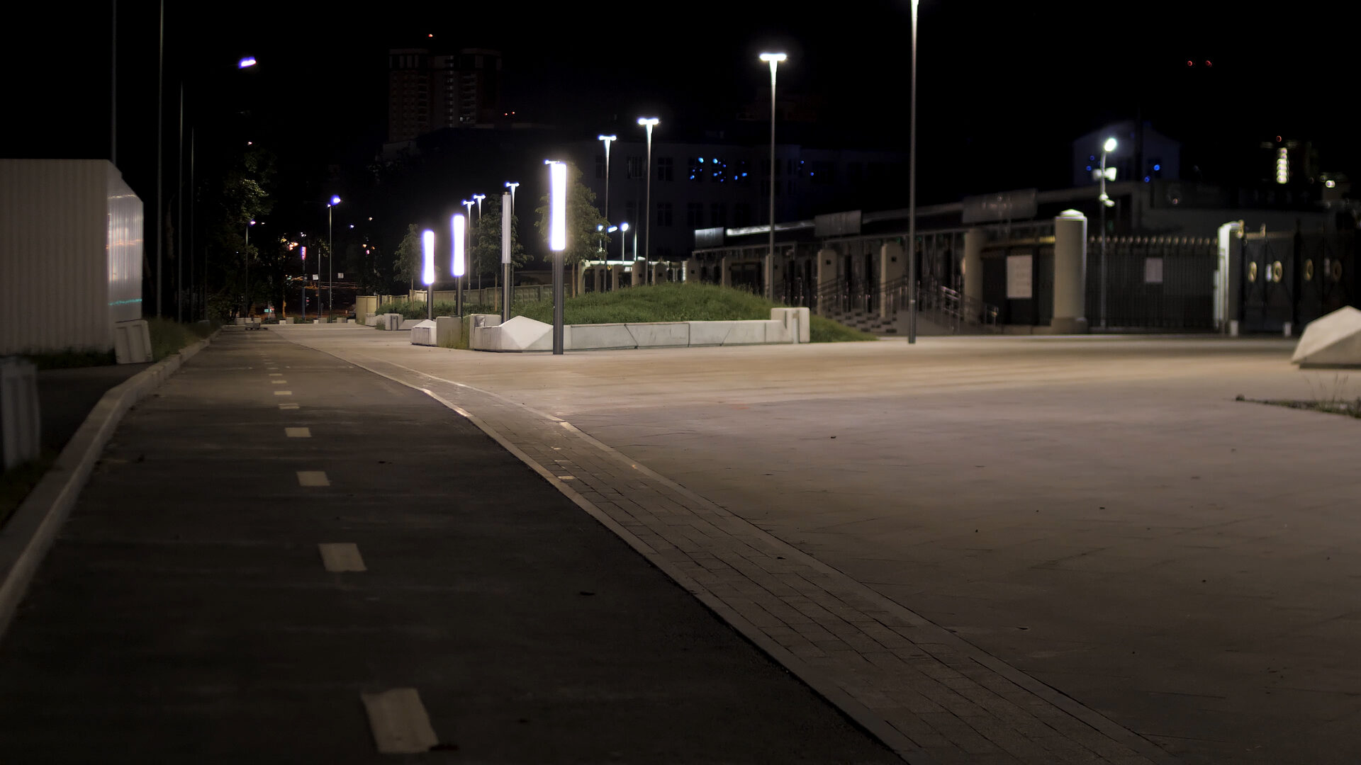
Nyctalopia is impaired vision under dim illumination or unusually slow dark adaptation. It differs from complete blindness at night. Patients may function in a well-lit room yet struggle outdoors after sunset, in a dark restaurant, on an unlit staircase or when oncoming headlights reduce contrast.
The complaint is relative. A person may not realize that family members see more in the same environment. Children may cling to a wall, hesitate on stairs or avoid dark play without naming the problem.
Night difficulty can reflect retinal rod dysfunction, optical scatter, reduced pupil response, uncorrected blur or impaired neural adaptation.
Tunnel vision is a lay description for constricted peripheral fields with a remaining central island. A person may read small letters yet miss side obstacles, door frames, people and traffic. It is a field problem, not simply low acuity.
True concentric constriction occurs in disorders such as advanced RP or glaucoma, but field patterns vary. Ring scotomas can expand inward and outward. Neurologic lesions produce hemianopic patterns, and functional visual symptoms may not follow anatomical rules.
Formal perimetry maps the defect. No home flashlight test can define it reliably.
Rod photoreceptors are highly sensitive in dim light and dominate peripheral night vision. Cones support daylight, color and fine central detail. In classic rod-cone dystrophy, rod dysfunction appears first, creating night blindness and midperipheral field loss; cones become involved later.
Cone-rod dystrophy reverses the emphasis, with central acuity, color and light sensitivity often affected earlier. Some stationary disorders impair night signaling without progressive degeneration.
Dark adaptation also requires the retinoid visual cycle in retinal pigment epithelium. Genetic or nutritional disruption can therefore impair night vision even when the fundus initially looks subtle.
RP is a group of inherited retinal diseases caused by many different genes and inheritance patterns. Common early features are night blindness and peripheral-field loss. Bone-spicule pigment, narrowed vessels and a pale optic disc may appear, but early disease can lack the classic triad.
Progression varies widely. Some people retain central vision for decades; others have early severe disease. The label RP does not identify the gene or exact prognosis.
Family history can be absent because of recessive inheritance, a new variant, small families, variable expression or unrecognized symptoms.
Congenital stationary night blindness produces lifelong dim-light difficulty that is often nonprogressive or slowly changing, with myopia, nystagmus or strabismus in some forms. The fundus may look normal. ERG patterns distinguish signaling defects.
Oguchi disease and fundus albipunctatus are other inherited night-vision phenotypes with characteristic examination or dark-adaptation findings. Some conditions once called stationary can have later changes.
Accurate molecular and electrophysiologic diagnosis prevents every child with night difficulty from being told they have progressive RP.
Vitamin A is essential to the visual cycle. Deficiency can cause night blindness and ocular-surface disease, sometimes with systemic malnutrition. Causes include severe dietary insufficiency, fat-malabsorption disorders, bariatric or intestinal surgery, pancreatic or liver disease and other medical conditions.
Testing and replacement require medical supervision. High-dose vitamin A can cause liver toxicity, birth defects and other harm. A patient should not self-treat night blindness with megadoses.
When deficiency is confirmed and treated early, visual function may improve, making it an important differential diagnosis.
Cataract scatters light, reduces contrast and creates glare or halos. Night driving can become difficult even while retinal rods function normally. A cloudy posterior subcapsular or cortical lens may be especially disruptive under glare.
Refraction, slit-lamp examination and glare history help distinguish optical degradation from retinal-field loss. Cataract surgery may improve contrast, but it cannot restore photoreceptors lost to RP.
Preoperative macular and retinal assessment sets realistic expectations.
Myopia, astigmatism and an outdated prescription reduce low-contrast clarity more in dim conditions. Larger pupils expose optical aberrations. Dry eye adds fluctuating scatter and glare.
These common problems can coexist with inherited retinal disease. Improvement with refraction or surface treatment does not necessarily explain persistent dark-adaptation delay or field loss.
A complete examination avoids both overdiagnosing dystrophy and attributing everything to glasses.
Glaucoma damages retinal ganglion cells and the optic nerve, causing peripheral-field loss that can progress toward a central island. Open-angle disease is often symptom-free until advanced. Night mobility may become difficult because field and contrast are reduced.
Pressure, corneal thickness, angle anatomy, optic-nerve appearance, OCT and visual fields are interpreted together. Normal pressure does not exclude glaucoma.
Glaucoma treatment lowers pressure; it differs from treatment and counseling for photoreceptor dystrophy.
A rhegmatogenous detachment often creates a sudden curtain or shadow, sometimes preceded by flashes and floaters. It is not “tunnel vision developing overnight from RP.” Urgent surgical evaluation protects remaining retina.
Tractional and exudative detachments have different mechanisms but can also reduce field. Pain is often absent.
Any abrupt monocular field loss, new flashes or shower of floaters bypasses routine inherited-disease scheduling.
Stroke, tumor, inflammation and other post-retinal lesions can produce hemianopia, quadrantanopia or other defects. Patients may call any side loss tunnel vision. Associated weakness, speech change, headache or sudden onset increases urgency.
Pupils, visual-field pattern, optic nerve and neurologic examination help localize the lesion. Brain imaging may be necessary.
An established retinal diagnosis does not protect a patient from stroke. New patterns require fresh evaluation.
Some medicines alter dark adaptation, pupil function or retina. Hydroxychloroquine toxicity typically affects parafoveal or pericentral retina rather than classic early night blindness. Retinoid drugs, phenothiazines and other agents can have ocular effects depending on dose and exposure.
A complete medication and supplement list is essential. Do not stop a necessary drug independently; the prescribing and eye clinicians coordinate risk and alternatives.
Herbal products can also contain vitamin A analogues or unknown ingredients.
Hearing loss raises concern for Usher syndrome in a person with rod-cone dystrophy. Balance problems, developmental differences, kidney disease, obesity, neurologic symptoms, skeletal findings or metabolic abnormalities may point toward other syndromic inherited retinal diseases.
Review should be specific rather than a frightening catalog. A molecular diagnosis can direct hearing, renal or other surveillance when the phenotype supports it.
The eye may be the first organ that reveals a broader condition.
Ask when dim-light difficulty began, whether it was lifelong or progressive, and whether side vision, color, glare or central detail changed. School, driving, falls and mobility examples are often more accurate than a severity number.
Family history includes vision, hearing, consanguinity and relatives with vague “night problems.” A three-generation pedigree can reveal dominant, recessive, X-linked or mitochondrial patterns without proving one.
Diet, intestinal surgery, liver disease, medications, trauma and neurologic symptoms identify acquired causes.
The clinician assesses acuity, color, pupils, pressure, lens, optic nerve, vessels, macula and peripheral retina. RP may show pigment migration, vessel attenuation and disc pallor, but appearance varies by gene and stage.
White dots, crystals, macular atrophy, schisis, flecks or normal-appearing retina can suggest other diagnoses. Cataract may obscure the view.
A clinical phenotype guides testing but should not be forced into a familiar label when findings conflict.
Automated static perimetry maps sensitivity at fixed locations; kinetic perimetry moves targets and can better characterize broad peripheral islands. Test choice depends on remaining field, age and ability.
Ring scotomas and progressive constriction are common in RP. Reliability indices, fixation, learning, fatigue and cataract affect results. Serial testing should use comparable methods.
Field tests document function, support mobility planning and may serve as trial outcomes; they do not name the causal gene.
Full-field ERG measures summed retinal electrical responses under dark- and light-adapted conditions. It separates rod and cone dysfunction and can detect widespread disease before obvious fundus change. Electrode and protocol standards matter.
ERG is not a direct acuity test or a brain scan. A severely reduced full-field response can coexist with useful central vision because a small foveal area contributes little to the summed signal.
Multifocal and pattern ERG answer different localized or ganglion-cell questions and are not interchangeable.
OCT shows photoreceptor layers, ellipsoid zone, macular edema, epiretinal membrane and atrophy. Preserved ellipsoid-zone width can help characterize central structural reserve. Cystoid macular edema is a treatable contributor to blur in some RP eyes.
Fundus autofluorescence maps RPE-related fluorophores. Hyperautofluorescent rings often border stressed retina, while hypoautofluorescence can reflect atrophy. Bright or dark color is not simply better or worse without context.
Serial imaging tracks structure but cannot by itself predict an individual’s exact functional timeline.
Panels, exome or genome approaches may identify pathogenic variants. Testing can confirm diagnosis, clarify inheritance, identify gene-specific therapy or trial eligibility and guide family counseling. It should use an appropriately certified laboratory and phenotype-informed interpretation.
A negative result does not disprove inherited disease; current methods miss some variants and genes. A variant of uncertain significance is not a molecular diagnosis and should not be used alone for predictive testing.
Genetic counseling before and after testing addresses expectations, family implications, privacy and reanalysis.
Most RP forms do not yet have an approved gene-specific treatment. Voretigene neparvovec is FDA-approved for confirmed biallelic RPE65 mutation-associated retinal dystrophy in patients with viable retinal cells. It is not a general RP injection.
Clinical trials study gene augmentation, editing, RNA, cells, optogenetics and neuroprotection. Eligibility depends on gene, stage, anatomy and protocol. Enrollment does not guarantee benefit.
Treatable complications such as cataract, macular edema and refractive error should be addressed even when degeneration itself lacks a cure.
Older vitamin A studies in RP have generated longstanding confusion, and current gene-specific risks, liver toxicity, pregnancy concerns and evolving evidence make self-treatment unsafe. High-dose vitamin E has also raised concern in historical data.
No supplement applies to every inherited retinal disease. ABCA4-related disease is a prominent reason to avoid excess vitamin A exposure without specialist direction.
Patients should bring every product to an inherited-retinal specialist rather than assume an “eye vitamin” is protective.
Orientation and mobility training teaches scanning, cane skills, navigation and safe travel. Better lighting, contrast, tactile markers, electronic accessibility and task organization help night and field limitations.
Referral should not wait until central acuity collapses. Early training allows practice while more vision remains. Driving counseling uses measured field, acuity, adaptation and local law.
Vocational and educational accommodations can preserve independence and employment.
Dark-adaptation tests measure how visual sensitivity recovers after exposure to light. Rod recovery follows a characteristic course, and delayed recovery can support retinal or visual-cycle dysfunction. Tests differ in stimulus location, wavelength and duration, so results need method-specific interpretation.
Routine clinics may not have formal dark adaptometry. History, ERG, fields and imaging often provide enough evidence to refer to an inherited-retinal center. A phone application in a dark room cannot reproduce calibrated retinal testing.
Cataract, pupil size and poor instruction can influence performance. The test supplements rather than replaces structural and electrophysiologic evaluation.
Cone involvement can reduce color discrimination, often later in rod-cone dystrophy and earlier in cone-rod disease. Plate tests screen common congenital color defects but may be insensitive to acquired retinal change. More quantitative arrangements or computer tests can add detail.
Contrast sensitivity captures difficulty seeing low-contrast objects, faces, curbs or signs despite reasonable high-contrast acuity. Cataract, glaucoma and retinal disease all affect it. This helps explain why a patient struggles at dusk while still reading a small black-on-white chart.
Functional testing should answer a clinical or rehabilitation question, not simply accumulate abnormal numbers.
Parents may notice that a child stays near walls, hesitates when lights dim, trips at dusk, avoids dark rooms or needs a hand long after others adapt. Teachers may report difficulty during assemblies or stage transitions. Nystagmus, high refractive error or developmental delay can provide additional clues.
Children adapt cleverly and may never complain. A comprehensive pediatric retinal evaluation uses age-appropriate fields, imaging and electrophysiology. Sedation is not automatically necessary for every test, but cooperation and protocol matter.
Families should avoid testing by hiding objects in darkness or scaring the child. Observe natural function and seek evaluation.
Some adults realize only after driving, parenting or comparing with a partner that they have always seen poorly at night. Lifelong stable symptoms may indicate a stationary inherited disorder rather than progressive RP. Others normalize slow progression until field loss becomes substantial.
Old school records, childhood photographs, prior prescriptions and family stories can reconstruct onset. ERG can distinguish rod-system patterns even when the fundus is subtle.
Late recognition does not prove late onset. It also does not remove the need to exclude cataract, deficiency and acquired disease that may now worsen the baseline.
Rare autoimmune retinal disorders can cause rapid photopsias, field loss, night difficulty, color change and reduced ERG responses, sometimes with initially mild fundus findings. Cancer-associated or melanoma-associated forms are important but uncommon.
Diagnosis requires specialist correlation among symptoms, examination, electrophysiology, imaging and systemic evaluation. Antiretinal antibodies alone can be nonspecific and should not establish the diagnosis in isolation.
A subacute course over weeks or months differs from many inherited dystrophies and can influence urgency. Immunosuppressive treatment carries substantial risk and requires a confident multidisciplinary plan.
Beyond vitamin A deficiency, some metabolic diseases, severe liver or pancreatic disorders and malabsorption can impair retinal function. Syphilis, inflammatory disease and other infections can mimic pigmentary degeneration. Medication toxicity can create widespread dysfunction.
Laboratory testing is guided by history and phenotype rather than ordering every possible panel. Nutritional status, surgery history, weight loss, gastrointestinal symptoms, exposure and systemic signs identify reasonable targets.
Calling every pigmentary retina “genetic RP” can miss reversible or systemically important causes. Conversely, a normal laboratory screen does not exclude inherited disease.
Visual-field constriction can occur in functional neurologic symptoms without structural retinal or brain disease. A tubular field that does not expand with test distance, inconsistent responses and mismatch with navigation may raise suspicion, but no single sign should be used to accuse a patient of fabrication.
Retinal disease, glaucoma and neurologic causes must be assessed first. Functional symptoms are real and can coexist with organic disease. Compassionate explanation and appropriate neurologic or psychological care are more useful than confrontation.
ERG and imaging can provide objective context, but they do not measure every aspect of visual processing.
Night driving requires peripheral detection, contrast, glare recovery, adaptation, attention and reaction time. A person with preserved central acuity may be unsafe if fields are severely constricted. Conversely, a diagnosis alone does not determine legal status.
Licensing standards vary by jurisdiction and may specify binocular or monocular field extent. Clinicians can counsel based on measured function and local law. Restricted daylight driving is not appropriate for every disease or every stage and should not be improvised.
Stopping driving can affect employment and identity. Transportation planning should begin before an emergency forces it.
Peripheral loss and dim-light difficulty increase collision and fall risk, especially when combined with neuropathy, hearing loss or balance problems. Even lighting, illuminated stair edges, high-contrast tape, reduced clutter and consistent furniture placement improve safety.
Night-lights should guide routes without producing disabling glare. Motion sensors can help, but abrupt bright light may slow readaptation. An occupational therapist can tailor changes to the actual field.
Family members should announce approach rather than appearing suddenly from the side and should avoid rearranging important objects without discussion.
Students may need seating, digital text, extra transition time between lighting environments, accessible diagrams and orientation support. Extended test time alone may not solve field scanning or glare.
Workers can use larger monitors, magnification, screen readers, task lighting, contrast controls and route training. Employers may provide reasonable accommodations under applicable law. Vocational rehabilitation helps match tools to essential tasks.
Disclosure decisions are personal and context-dependent. A rehabilitation professional can help communicate functional needs without reducing the person to a diagnosis.
Posterior subcapsular cataract is common in RP and can worsen glare, contrast and central acuity. Surgery may provide meaningful benefit when the lens contributes substantially, but macular structure and field limit the ceiling. Preoperative OCT checks ellipsoid-zone reserve, edema and epiretinal membrane.
Inflammation, cystoid edema, capsule contraction and later posterior-capsule opacity require attention. Intraocular-lens choice should prioritize optical quality; premium lenses that reduce contrast may be unsuitable.
Improvement in acuity after surgery does not widen a constricted retinal field, so expectations must separate central clarity from peripheral function.
Fluid-filled cystic spaces can reduce central vision in some RP eyes. OCT detects them even when examination is subtle. Carbonic-anhydrase inhibitors, steroid or other treatments may be considered, with response and side effects monitored.
A drier OCT does not reverse peripheral rod loss, and treatment burden should be proportional to functional benefit. Recurrence is possible.
This treatable complication illustrates why regular examination remains useful even when no cure exists for the underlying gene disorder.
Usher syndrome combines inherited hearing loss with RP, with balance involvement in some types. Hearing differences may be congenital or progressive. Early recognition supports audiology, communication access, vestibular care and genetic counseling.
The visual and auditory burdens interact: a person who depends on lip reading may struggle as fields narrow, while someone using sign language needs adequate visual access. Rehabilitation should integrate both senses.
Genetic testing clarifies subtype and family risk but should be interpreted with the phenotype and counseling.
Dominant, recessive, X-linked and mitochondrial inheritance create different recurrence patterns. Affected siblings with unaffected parents may suggest recessive disease; only males in a maternal line can suggest X-linked inheritance, but real pedigrees are often incomplete.
Genetic counseling explains probabilities after molecular and family information is available. It supports—not directs—choices about partner testing, pregnancy, donor gametes or preimplantation testing.
Relatives should not receive predictive testing from a proband’s uncertain variant. A confirmed pathogenic finding is interpreted within the appropriate inheritance model.
Inherited-retinal trials may target a gene, mutation class, retinal cell, stage or functional endpoint. Screening includes molecular confirmation, OCT evidence of viable tissue, age and prior treatment. A trial listing is not proof that an intervention works.
Participants should understand randomization, sham procedures, travel, long follow-up, costs, alternatives and whether treatment could affect later eligibility. Registries can support research without providing therapy.
Commercial clinics that offer unapproved stem cells or gene products outside regulated trials can cause severe harm. Independent specialist review is essential.
Fields, ellipsoid-zone width, autofluorescence area, ERG and patient-reported function capture different aspects of disease. Change may be slow and nonlinear. Test variability makes short-term prediction difficult.
Gene and variant can inform natural history, but even relatives may progress differently. Clinicians should provide ranges and update counseling from serial data rather than announce an exact age of blindness.
Planning for accessibility is prudent without assuming that all useful central vision will disappear on schedule.
Night and field loss can produce anxiety, grief, social withdrawal and fear of independence. Children may hide difficulty to avoid being different; adults may delay mobility aids because they symbolize loss.
Peer support, counseling and rehabilitation can restore agency. A cane or screen reader is a tool, not evidence that vision has “failed.” Mental-health symptoms deserve direct care.
NRT stress support may complement these services when it does not replace evidence-based counseling or create false restoration promises.
More light is not always better. Even, shielded illumination can improve contrast, while a bare bright bulb creates glare and deep shadows that make adaptation harder. Task lights should be positioned to illuminate work without shining into the eyes. Warm and cool sources can be compared for comfort, but no household wavelength regenerates rods.
Carry a small flashlight or use an accessible phone-light shortcut for transitions, keeping hands available for railings. Give the eyes time to adapt when moving between bright and dark areas. Tinted lenses may reduce photophobia outdoors but can worsen dim-light function if worn inappropriately.
A low-vision professional can test filters, contrast and placement in the actual task. Product advertisements that promise “night-vision restoration” should be judged against measured fields and dark-adaptation outcomes, not bright-room demonstrations.
Record real tasks rather than rating vision abstractly: time needed to adapt after entering a theater, missed objects from the side, falls, reading endurance, glare and which eye seems different. Note whether change was sudden or gradual and any hearing, neurologic, dietary or medication context.
The diary helps clinicians select tests and helps rehabilitation target safety. It should not become repeated self-testing that increases anxiety. Sudden curtain, flashes, floaters or neurologic change bypasses the diary and receives urgent care.
NRT may support sleep, stress, balanced nutrition, activity, coping and adherence while inherited-retinal evaluation and rehabilitation continue. These goals can improve quality of life.
NRT cannot regenerate rods or cones, widen a genetically constricted field, replace RPE65 gene therapy, treat a detachment or correct vitamin deficiency without medical diagnosis. Subjective ease in dim light is not proof of restored photoreceptors.
Herbs and supplements can cause toxicity or interaction. Acupuncture must not delay urgent evaluation of sudden field loss.
Learn about retinitis pigmentosa and Netra Restoration Therapy, review Netra Eye Institute’s approach, read about living well with low vision, or request an appointment.
No. Older eyes need more light, but substantial dim-light difficulty can reflect cataract, retinal or systemic disease and deserves evaluation.
No. Glaucoma, neurologic disease, retinal detachment and other conditions can constrict or remove peripheral field.
Central acuity may remain excellent. Formal visual fields evaluate peripheral function.
It can treat confirmed deficiency, but high-dose use without diagnosis can be toxic and may be inappropriate in inherited disease.
No established evidence shows regeneration of lost photoreceptors or reliable field expansion. NRT is supportive only.
Night blindness and tunnel vision are functional clues, not disease names. Their timing and pattern direct an evaluation across retina, lens, optic nerve, brain, nutrition and genetics.
Gradual inherited disease needs precise phenotyping, counseling and rehabilitation; sudden field loss needs emergency care. NRT may support adaptation and health, never replace those pathways.
Medical Disclaimer: This article provides general education and is not medical advice, diagnosis or a recommendation for vitamins, herbs, gene therapy or NRT. Sudden field loss, a curtain, flashes, new floaters, major blur, eye pain, redness, weakness, speech change or severe headache requires urgent care. Do not take high-dose vitamin A or change prescribed medicines without responsible clinicians. Netra Restoration Therapy is adjunctive and cannot replace dilation, visual fields, OCT, autofluorescence, ERG, genetic testing, retinal surgery, glaucoma treatment, deficiency evaluation, rehabilitation or emergency care.



