
Genetic counseling visit for an inherited eye condition

Clinical guide

Wolfram Syndrome and Vision Loss: What Families Should Know

How WFS1-related optic atrophy develops, what monitoring should track, and where Netra Restoration Therapy fits as adjunctive supportive care in a Wolfram syndrome comanagement plan.

September 1, 2026

Wolfram syndrome affects the eye through optic atrophy: the optic nerve thins, usually beginning in childhood or early adulthood, causing gradual loss of color vision, central detail and visual field. It is caused by variants in the WFS1 gene. This page explains what happens to vision, how it is monitored, and what supportive care involves.

An integrative perspective on WFS1-related optic neuropathy and coordinated supportive care.

By Saikumar Gandapodi DAOM, L.Ac., Dipl. OM.

Printable edition. This article is also available as a designed one-page PDF: Wolfram Syndrome: Mechanisms, Function, and NRT Support (PDF).

Wolfram syndrome is a rare, progressive, multi-system neurodegenerative disorder caused by biallelic WFS1 (or, less commonly, CISD2) variants. Optic atrophy is a defining feature, and the axonal loss is irreversible. Netra Restoration Therapy (NRT) may be incorporated into a comanagement plan as adjunctive supportive care alongside, and never in place of, genetic, neuro-ophthalmic, endocrine, audiologic and neurologic evaluation and treatment.

How Wolfram Optic Atrophy Develops

Wolframin is an endoplasmic reticulum (ER) membrane protein. Its loss produces a persistent unfolded-protein response and, critically, depletion of ER calcium stores through reduced SERCA2 activity and ryanodine receptor leak. Impaired IP3R–GRP75–VDAC transfer at mitochondria-associated ER membranes then lowers mitochondrial matrix calcium, pyruvate dehydrogenase activation and ATP output. Retinal ganglion cells are unusually exposed to this deficit because their long axons demand continuous energy for conduction and transport.1,2

An Axon-First Degeneration

Wolfram optic atrophy does not begin at the cell body. WFS1-deficient optic nerves show synaptic change, myelin disorganization and reduced oligodendroglial MCT1/basigin-dependent lactate delivery before substantial ganglion-cell loss. Comparative OCT work found peripapillary RNFL thinning appearing early while ganglion-cell-layer thinning emerged considerably later. This dying-back pattern distinguishes Wolfram syndrome from OPA1-related and other primary mitochondrial optic neuropathies, and it means structural monitoring should not wait for acuity to decline.3,4

Multisystem Context and Modifiers

Optic atrophy rarely presents in isolation. Juvenile-onset diabetes mellitus usually precedes it, and diabetes insipidus, sensorineural hearing loss, urinary-tract dysfunction, ataxia, autonomic instability and psychiatric manifestations may follow. Glycemic instability, sleep disruption, nutrition, mood and treatment burden can all amplify functional disability. These shape how a patient experiences the disease; they are not the cause of the optic neuropathy.5,6

Where NRT Fits in a Comanagement Plan

NRT is positioned as adjunctive supportive care around the established medical plan.

  • Constituent-level ER-stress mitigation in experimental models: astragaloside IV (AS-IV) restores SERCA activity in non-ocular, non-Wolfram systems; it does not restore wolframin function.1,9,10
  • Mitochondrial resilience: mitochondrial dysfunction is established in Wolfram neurons. Danshen constituents have supported calcium handling and redox endpoints in non-Wolfram injury models.11,12
  • RGC stress resistance: AS-IV and tanshinone IIA have protected retinal ganglion cells in acute traumatic and ischemic injury models; none were Wolfram models.13,14
  • Ocular-surface support: address dry eye, glare and photophobia that compound the functional impact of reduced acuity and contrast.
  • Glial, neuro-inflammatory and trophic support: salvianolic acid B acts through Nrf2-dependent glial protection in neurotoxin models, and Astragalus preparations have influenced NGF signaling in optic nerve injury.15,16
  • Metabolic and lifestyle support: sleep, stress physiology, activity and nutrition, coordinated with endocrinology-directed glycemic management.
  • Whole-person care: attention to fatigue, mood, anxiety and caregiver burden, in coordination with appropriate low-vision rehabilitation specialists.

Diagnosis Is Genetic and Multimodal

Molecular confirmation is central. Biallelic WFS1 or CISD2 testing, with genetic counseling, should be pursued in any young patient with unexplained bilateral optic atrophy and diabetes mellitus, because monoallelic dominant WFS1-spectrum disease differs in inheritance and course. Structural and functional assessment should include serial peripapillary RNFL and macular ganglion-cell OCT, automated perimetry, color vision and contrast sensitivity, with pattern ERG or VEP where informative, and MRI when anatomic spread is in question.3,5,7

Coordinated Care

Care is inherently multidisciplinary: neuro-ophthalmology, endocrinology, medical genetics, audiology, urology, neurology, psychiatry and low-vision rehabilitation. There is no approved disease-modifying therapy. Investigational directions include calcium-stabilizing repurposing candidates and RGC-directed WFS1 gene delivery, which preserved visual and axonal outcomes in a WFS1-null mouse.2,8

Boundaries of NRT in Wolfram Syndrome

  • It does not correct the underlying WFS1 or CISD2 genetic lesion.
  • It does not regenerate lost RGC axons or restore vision already lost.
  • It must never delay genetic confirmation, endocrine and neurologic management, or surveillance by a neuro-ophthalmologist.
  • Herbal constituents may interact with anticoagulants and with insulin or oral hypoglycemics; medication review with the treating clinician is required before starting.17

Measuring Potential Benefit

Because the disease is slowly progressive, short-term symptom change is not a valid measure of benefit. Anchor assessment to serial structure and function: RNFL and RGC on OCT, perimetric mean deviation, contrast sensitivity, color vision and electrophysiology where available. Track patient-centered outcomes separately — reading and mobility function, glare, fatigue, sleep, mood and quality-of-life measures — and interpret any change against documented natural history rather than expectation.3,4

The therapeutic target for NRT in Wolfram syndrome is not vision alone: it is comfort, functional capacity, systemic stability and quality of life, delivered around an established medical plan.
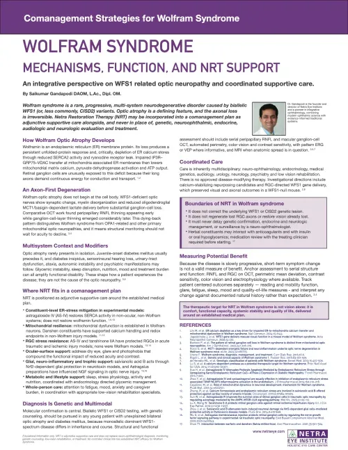
Download the Article (PDF)

Comanagement Strategies for Wolfram Syndrome article cover

Download: Wolfram Syndrome — Mechanisms, Function, and NRT Support (PDF)

References

  1. Liiv M, et al. ER calcium depletion as a key driver for impaired ER-to-mitochondria calcium transfer and mitochondrial dysfunction in Wolfram syndrome. Nat Commun. 2024;15:6143.
  2. Jagodzinska J, et al. WFS1 gene delivery rescues visual function in a mouse model of Wolfram syndrome. Acta Neuropathol Commun. 2026;14:120.
  3. Barboni P, et al. The pattern of retinal ganglion cell loss in Wolfram syndrome is distinct from mitochondrial optic neuropathies. Am J Ophthalmol. 2022;241:206-216.
  4. Rossi G, et al. MCT1-dependent energetic failure and neuroinflammation underlie optic nerve degeneration in Wolfram syndrome mice. eLife. 2023;12:e81779.
  5. Urano F. Wolfram syndrome: diagnosis, management, and treatment. Curr Diab Rep. 2016;16:6.
  6. Rigoli L, et al. Genetic and clinical aspects of Wolfram syndrome 1. Pediatr Res. 2018;83:921-929.
  7. de Heredia ML, et al. Genotypic classification of patients with Wolfram syndrome. Genet Med. 2013;15:497-506.
  8. Lu S, et al. A calcium-dependent protease as a potential therapeutic target for Wolfram syndrome. Proc Natl Acad Sci USA. 2014;111:E5292-E5301.
  9. Guo H, et al. Astragaloside IV attenuates podocyte apoptosis mediated by endoplasmic reticulum stress through upregulating sarco/endoplasmic reticulum Ca2+-ATPase 2 expression in diabetic nephropathy. Front Pharmacol. 2016;7:500.
  10. Zhao Y, et al. Astragaloside IV and cycloastragenol are equally effective in inhibition of endoplasmic reticulum stress-associated TXNIP/NLRP3 inflammasome activation in the endothelium. J Ethnopharmacol. 2015;169:210-218.
  11. Cagalinec M, et al. Role of mitochondrial dynamics in neuronal development: mechanism for Wolfram syndrome. PLoS Biol. 2016;14:e1002511.
  12. Zhang JY, et al. Calcium homeostasis and endoplasmic reticulum stress are involved in salvianolic acid B-offered protection against cardiac toxicity of arsenic trioxide. Oncotarget. 2017;8:97384-97393.
  13. Sun W, et al. Astragaloside IV improves the survival rates of retinal ganglion cells in traumatic optic neuropathy by regulating autophagy mediated by the AMPK-MTOR-ULK signaling pathway. Mol Vis. 2025;31:99-112.
  14. Lu X, Wang W. Tanshinone II-A protects retinal ganglion cells against retinal ischemia/reperfusion injury. Int J Clin Exp Pathol. 2016;9:11230-11237.
  15. Zhou J, et al. Salvianolic acid B attenuates toxin-induced neuronal damage via Nrf2-dependent glial cells-mediated protective activity in Parkinson's disease models. PLoS One. 2014;9:e101668.
  16. Wu Q, et al. Astragalus membranaceus injection protects retinal ganglion cells by regulating the nerve growth factor signaling pathway in experimental rat traumatic optic neuropathy. Evid Based Complement Alternat Med. 2020;2020:2429843.
  17. Chan TY. Interaction between warfarin and danshen (Salvia miltiorrhiza). Ann Pharmacother. 2001;35:501-504.
Educational information only. NRT is adjunctive supportive care and does not replace neuro-ophthalmologist diagnosis, monitoring, genetic counseling, low-vision rehabilitation, or treatment. No controlled clinical trial has established NRT efficacy for Wolfram syndrome.

More on inherited retinal disease

Request Consultation
Conditions We Treat