
Search conditions, treatments, research reviews and articles. Press Enter to see all results, Esc to close.
Search conditions, treatments, research reviews and articles. Press Enter to see all results, Esc to close.
Blog
October 31, 2026
A diabetic tractional retinal detachment (TRD) happens when scar tissue from proliferative diabetic retinopathy shrinks and pulls the retina away from the back of the eye [1]. If the detachment spares the macula, the center of the retina, and is not growing, it can sometimes be watched closely. If it involves or threatens the macula, or a tear forms as well, vitrectomy surgery is usually advised as soon as possible [2].
This article is general education. It does not replace advice from your own retina specialist.
Key takeaways
In this article
Go to an eye doctor or emergency room right away if you have a new curtain or shadow, flashes of light, a sudden shower of floaters, or a sudden drop in vision. If you already know you have a tractional detachment, call your retina specialist the same day about any new blur or distortion in your central vision [6,2].
Do not wait for a routine appointment. Our guide to urgent vision changes explains other eye symptoms that need same-day care.
It is a detachment caused by pulling, not by a tear. In proliferative diabetic retinopathy, abnormal new vessels grow with scar tissue on the retina's surface. As that tissue slowly contracts, it lifts the retina off the back of the eye. The detached area is usually firm, does not move, and has no breaks [1,2].
The retina is the light-sensing layer at the back of the eye. There are three main kinds of retinal detachment: rhegmatogenous (caused by a tear or hole), tractional (caused by pulling) and exudative (caused by fluid leaking under the retina) [10]. Diabetic eyes can develop a tear next to scar tissue. This is a combined tractional and rhegmatogenous detachment, and it can worsen quickly [2].
TRD is a late complication of diabetic retinopathy. A dilated eye exam at least once a year and good diabetes care help prevent or delay vision loss [1]. Learn how the disease advances in the four stages of diabetic retinopathy.
Symptoms depend on where the retina is pulled. You may notice blurred or distorted central vision or a missing area in part of your vision. Floaters, cobwebs or a dark haze usually mean bleeding. A sudden curtain, flashes or a burst of floaters can mean a tear has formed as well [2,8,6].
Because traction builds gradually, a detachment away from the macula may cause little change in central vision at first. Central vision usually drops sharply once the macula is involved [2]. That is why your retina specialist may find a TRD at a routine visit before you notice it.
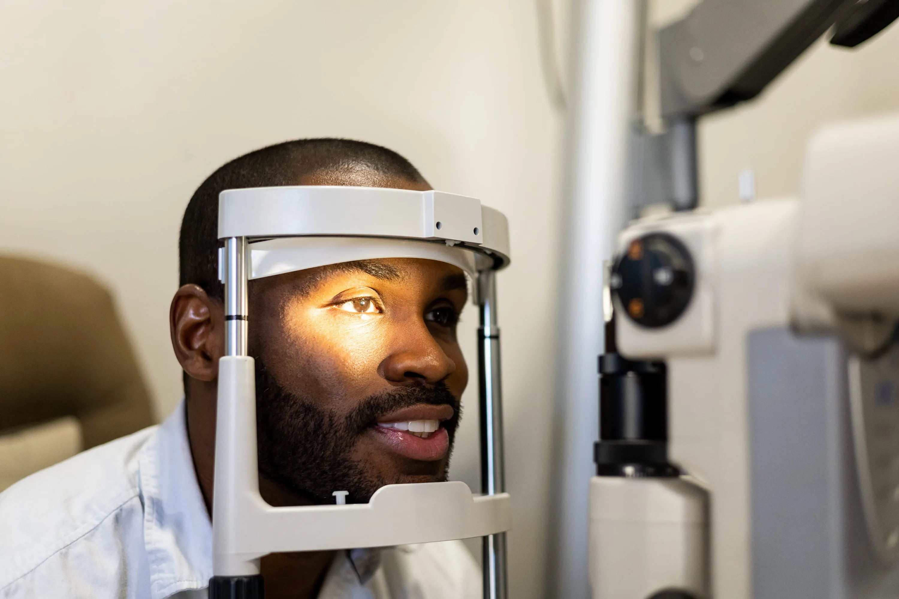
To see the detachment, the doctor uses a dilated exam and an OCT scan of the macula. If blood blocks the view, a B-scan ultrasound can show traction and detachment behind the blood [2]. Our guide to diabetic eye exams and imaging explains these tests.
The macula gives you sharp central vision. If a tractional detachment spares it (macula-on) and is small and stable, close monitoring may be reasonable. If the macula is detached (macula-off) or threatened, vitrectomy is usually advised as soon as possible, because a detached macula can lose vision permanently [2,10].
| Situation | What it means | Usual plan | Outlook |
|---|---|---|---|
| Macula-on, small and stable | Traction is away from the center; central vision is still attached | Close checks with OCT; laser for active disease; surgery if it grows [2] | Best chance to keep central vision if treated before the macula detaches [10] |
| Macula threatened | Traction is spreading toward the center | Vitrectomy is often advised before the macula detaches [2] | Depends on how soon the pull is released |
| Macula-off | The center of the retina has lifted | Vitrectomy as soon as possible [2] | Macular detachment predicted poorer final vision in a 2025 study [11] |
| Combined tractional and rhegmatogenous | A tear lets fluid under the retina as well | Vitrectomy as soon as possible; can worsen quickly [2] | More complex; in one study, eyes needing silicone oil were more often combined detachments and had lower success [4] |
| Macula-off for more than 6 months | Long-standing detachment | Surgery may still help selected eyes [12,13] | Guarded; depends on blood flow and starting vision [12] |
In a 1981 study of 99 eyes with traction outside the macula, about 14% went on to macular detachment within a year without surgery [3]. A 2024 review notes that small detachments outside the center can stay stable for years after laser [2]. Observation needs a plan: a set check-up interval, repeat scans, and clear signs that would lead to surgery.
Ask what change would make your surgeon recommend an operation. Common triggers are growth toward the macula, new bleeding that hides the view, or a new tear [2].
The main treatment is pars plana vitrectomy. The surgeon removes the vitreous gel, cuts and peels the scar tissue to release the pull, and applies laser to the retina. A gas bubble or silicone oil may be left in the eye to hold the retina in place while it heals [2,14].
Laser and anti-VEGF injections treat the active blood vessels. They do not release scar tissue that is already pulling on the macula, so detachments that involve or threaten the center usually need surgery [2]. Our guide to injections, laser and surgery compares these treatments.
Many surgeons inject an anti-VEGF drug a few days before vitrectomy to shrink fragile vessels [2]. A 2023 Cochrane review of 28 trials (1,914 eyes) found it probably reduced early bleeding after surgery, from 31% to 12%. It also cut retinal breaks during surgery from 31% to 12%, and it may improve vision [15].
Timing matters. A 2026 meta-analysis found the lowest bleeding rates when the injection came 1 to 7 days before surgery, but it could not name one best interval [16]. In eyes with heavy scarring, the injection can make scar tissue contract. In one series of eyes with severe disease, a TRD developed or worsened after 11 of 211 injections (5.2%), on average 13 days later [17].
This does not mean injections commonly cause TRD. A pooled analysis of five DRCR trials, in eyes where surgery was not planned, found no higher TRD risk with anti-VEGF than with laser, sham or saline treatment [18]. Ask what will happen if your operation is delayed after an injection [2].
Reattachment rates are high. In a study of 403 eyes, the retina was reattached in 87.6% after one surgery and 92.6% by final follow-up. Vision improved by 2 or more lines in 56.3%, stayed stable in 23.8% and got worse in 19.9%. A 2024 meta-analysis pooled reattachment at about 95% [4,5].
Reattachment and vision are different outcomes. The retina can be back in place while vision stays limited by damage to the macula or poor blood flow [12]. The meta-analysis also found signs of publication bias, so the average results may be somewhat optimistic [5].
Factors linked to poorer vision after surgery include:
Sometimes. A 2025 study looked at 175 people with very poor vision and a macula-off detachment for more than 6 months. Those who had vitrectomy gained far more vision than those who declined it. They also had fewer cases of total vision loss and neovascular glaucoma. The study was not randomized [13].
A 2016 study of 96 eyes with macular detachment for at least 6 months found the retina reattached in 90.6% after one surgery. The share of eyes seeing 20/200 or better rose from 17.7% to 41.6% [12]. These results support a careful discussion with a surgeon. They are not a reason to delay treatment of a new or growing detachment.
Most people go home the same day. The eye can stay red and blurry for several weeks, and many people take 2 to 4 weeks off work. With a gas bubble, you may need to hold a head position and must not fly or travel to high altitude until it is gone [14].
If silicone oil is used, you may need a second operation to remove it [14].
If some vision loss remains, low-vision rehabilitation can help with reading, getting around and managing medicines. For a broader overview, see our diabetic retinopathy guide.
Netra Restoration Therapy (NRT) is Netra Eye Institute's integrative program based on acupuncture and Oriental medicine, offered alongside care from eye doctors. NRT cannot release scar tissue, reattach the retina, or stop a detachment from growing. It cannot replace laser, injections or vitrectomy.
NRT has not been tested in controlled trials for tractional retinal detachment. Never delay an exam or surgery because of NRT; a detachment that involves or threatens the macula needs prompt retinal care. If you are curious about NRT, raise it with your retina specialist first. You can read what NRT involves and what is known about its effectiveness.
It depends on the macula and on whether a tear is present. A macula-off or combined detachment needs vitrectomy as soon as possible. A small, stable detachment away from the macula is not an emergency but needs close follow-up. Any sudden curtain, flashes or vision loss is an emergency [2,6].
Sometimes, if it is small and away from the macula. Laser to the retina can calm the disease, and some of these detachments stay stable for years with close checks. Detachments that involve or threaten the macula usually need vitrectomy [2,3].
A tractional detachment is caused by scar tissue pulling the retina, usually without a tear. A rhegmatogenous detachment starts with a tear or hole that lets fluid get under the retina. Diabetic eyes can have both at once, which tends to worsen quickly [10,2].
Macula-off means the macula, the part of the retina used for reading and seeing faces, has lifted away from the back of the eye. Central vision usually drops sharply. A detached macula can lose vision permanently, so surgery is advised as soon as possible [10,2].
In a study of 403 eyes, the retina was reattached in 87.6% after one operation and 92.6% by final follow-up. Vision improved in 56.3% and stayed stable in another 23.8% [4].
Uncommonly, and mainly in eyes with heavy scarring. In one series of eyes with severe disease, a TRD developed or worsened after 11 of 211 bevacizumab injections, about 2 weeks later on average. A pooled analysis of five DRCR trials found no higher risk overall, so surgeons time pre-surgery injections close to the operation [17,18,2].
Expect weeks to months. The eye is often red and blurry for several weeks, many people need 2 to 4 weeks off work, and a gas bubble takes time to fade. Silicone oil removal adds another operation [14].
Often not fully. Surgery reattaches the retina in most eyes, but vision depends on macular damage, blood flow in the macula and how severe the detachment was. In one large study, about 1 in 5 eyes lost 2 or more lines of vision despite surgery [4,11,12].
Sources checked October 31, 2026.
This article provides general education and does not replace an individual eye examination, diagnosis or treatment plan. A new curtain or shadow, flashes, a sudden shower of floaters or a sudden drop in vision can mean retinal detachment; go to an eye doctor or emergency room right away.