
Search conditions, treatments, research reviews and articles. Press Enter to see all results, Esc to close.
Search conditions, treatments, research reviews and articles. Press Enter to see all results, Esc to close.

Clinical guide
What the research on inflammation in Fuchs endothelial corneal dystrophy shows and does not show, how the disease is monitored, and where Netra Restoration Therapy fits as adjunctive support alongside the corneal specialist.
October 17, 2026
Fuchs endothelial corneal dystrophy (FECD) slowly damages the cornea's inner cell layer, causing morning blur, glare and, in time, persistent clouding. It is inherited and degenerative, but studies of the fluid inside the eye also show a quiet, low-grade inflammation that grows as the disease advances. This page explains what that research shows, how the disease is monitored, and where supportive care fits.
An integrative perspective on endothelial failure, inflammation and coordinated supportive care.
By Saikumar Gandapodi DAOM, L.Ac., Dipl. OM.
Printable edition. This article is also available as a designed one-page PDF: Fuchs Endothelial Corneal Dystrophy: Mechanisms, Inflammation, and NRT Support (PDF).
Many people with Fuchs dystrophy first notice it in the morning. Vision is cloudy on waking and clears over a few hours. Then one day it doesn't fully clear. By the time they see a corneal specialist, they have usually heard two things: that the condition is inherited, and that a corneal transplant may eventually be needed.
Both statements are true. But they leave out part of the picture. Over the past decade, researchers sampling the fluid inside the eye have repeatedly found signs of a slow, smoldering inflammation, even in eyes that look perfectly calm on examination. What follows explains what those studies found, what they do not prove, and how that knowledge shapes the way we coordinate care.
Fuchs dystrophy is not caused by inflammation. It is driven mainly by genetic susceptibility, cellular stress and aging in a layer of cells that cannot replace itself. Inflammation appears to be a secondary amplifier: it follows the damage and may help it progress. When vision fails, endothelial keratoplasty performed by a corneal surgeon remains the established treatment. Integrative care, including Netra Restoration Therapy (NRT), can support the eye around that plan. It does not replace it.
The cornea is the clear front window of the eye. Its innermost layer, the endothelium, is a single sheet of cells that constantly pumps fluid out of the cornea to keep it thin and transparent. Adult endothelial cells barely divide in the living eye, so cells that are lost are not replaced; neighboring cells spread out to cover the gap.
In Fuchs dystrophy, these cells are lost faster than normal. Small collagen bumps called guttae form on the membrane the cells sit on (Descemet membrane). As pumping capacity falls, the cornea holds more water. Late-onset Fuchs dystrophy affects roughly 4% of people over 40 in Western populations, is more common in women, and is one of the most common reasons for corneal transplantation.
Dry eye and cataract often coexist and can add their own blur, so a careful examination matters.
Fuchs dystrophy does not look like an inflamed eye. There are usually no inflammatory cells, no flare and no redness. Yet when researchers sample the aqueous humor, the clear fluid that bathes the endothelium, they repeatedly find altered immune signals. The pattern is a slow, chronic one: chemical messengers that attract immune cells (chemokines), activation of the complement system and fibrosis-promoting factors. The sudden, acute signals typical of uveitis are usually not raised.
The signal looks different at different stages:
A 2026 laboratory study adds a mechanism. Endothelial cells exposed to UVA light and an oxidized estrogen metabolite entered senescence, a state of permanent arrest in which cells release inflammatory signals. They produced IL-8 and IL-17 and laid down scar-forming proteins. Blocking those signals, or clearing the senescent cells, reduced the scarring [11]. This links oxidative stress, the higher rate in women, inflammation and the scar layer seen in advanced Fuchs dystrophy.
A reasonable working model is a feed-forward loop. Stressed endothelial cells release danger signals, the eye's normal immune protection weakens, and chemokines, complement and TGF-β encourage further cell loss and scarring. Breaking that loop is an active research goal.
Many people with Fuchs dystrophy also need cataract surgery. Studies show that inflammatory and scar-promoting signals in the eye can stay raised for at least three months afterward, and that higher TGF-β and MCP-1 levels go along with more corneal swelling [9, 10]. This is one reason corneal surgeons think carefully about whether to combine cataract surgery with a transplant or stage them. That decision belongs to the surgeon. Integrative support should be planned around it.
Because the cornea is thickest after a night with the eyes closed, measurements should be repeated at a similar time of day.
Hypertonic (salty) drops and ointments are sometimes used for morning blur. In a randomized trial, however, hyperosmolar drops did not clear morning swelling faster and caused more irritation [15]. When the endothelium can no longer keep the cornea clear, a corneal surgeon replaces it with donor tissue through Descemet membrane endothelial keratoplasty (DMEK) or Descemet stripping automated endothelial keratoplasty (DSAEK). Newer approaches are being studied, but transplant remains the definitive treatment for advanced disease.
NRT is an integrative program that combines evidence-informed herbal medicine, acupuncture, nutrition and whole-person care. In Fuchs dystrophy we use it as adjunctive support around the corneal specialist's plan. Its goals follow the biology described above:
These laboratory findings give a reason to investigate. They are not proof that NRT works for Fuchs dystrophy. Patients have reported improvements in comfort and vision, but whether NRT changes inflammatory markers inside the eye or slows the disease still needs clinical study.
Netra Eye Institute works alongside your corneal specialist, who leads staging, monitoring and the timing of cataract surgery and transplant. With your consent, both teams share examination and imaging results, the NRT plan and any herbs or supplements, and agree on follow-up. We report changes in your vision or comfort and refer you back promptly for worsening swelling, pain or vision loss. Around surgery, NRT is paused or adjusted on the surgeon's advice.
We track visual acuity, contrast and glare, how long morning blur lasts, daily activities such as reading and driving, corneal thickness and tomographic swelling, measured at a consistent time after waking. Endothelial imaging is repeated on the same device where possible. A higher cell count on its own does not prove regeneration, because sampling and image quality vary.
The therapeutic target for NRT in Fuchs dystrophy is not endothelial regeneration: it is to reduce degeneration, improve usable vision and comfort, support a calmer environment inside the eye, and keep daily life going, with timely access to corneal treatment when it is needed.
Not primarily. It is a genetic and degenerative disease of the corneal endothelium. Research shows low-grade inflammation inside the eye that seems to follow the damage and may speed it up, especially in advanced disease and after cataract surgery.
There is no evidence that steroid or other anti-inflammatory drops stop guttae from forming or cells from being lost. Your surgeon may use them around surgery for other reasons.
Cataract surgery stresses the endothelium, and studies show inflammatory signals can remain raised for months afterward. Many people with Fuchs dystrophy still have successful cataract surgery. Your corneal surgeon will decide whether to combine or stage it with a transplant.
No. NRT does not remove guttae or regrow endothelial cells. It aims to support comfort, usable vision and a calmer environment inside the eye alongside your specialist's care. If your surgeon recommends a transplant, it should not be delayed.
Protect your eyes from UV light with good sunglasses, avoid smoking, keep blood sugar and general health in check, and treat dry eye. Tell your eye doctors about every supplement you take.
Often, yes, but timing matters. We coordinate with your surgeon and pause or adjust NRT around the procedure on their advice.
If you or someone you care for has Fuchs dystrophy, we can review your records, talk through how adjunctive care might fit alongside your corneal specialist's plan, and set realistic goals together. Request a consultation, call (732) 503-9999, or read more about Fuchs dystrophy and Netra Restoration Therapy. Eye care professionals can also visit our referring doctors page.
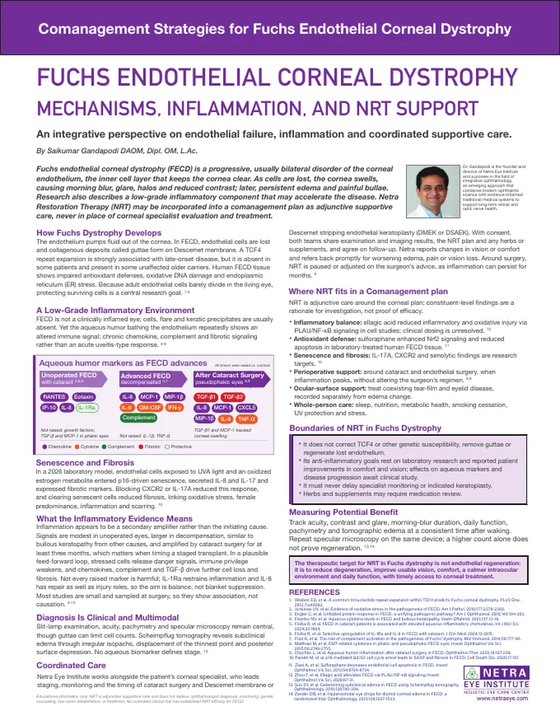
Download: Fuchs Endothelial Corneal Dystrophy — Mechanisms, Inflammation, and NRT Support (PDF)
Educational information only. NRT is adjunctive supportive care and does not replace diagnosis, monitoring or treatment by an ophthalmologist or corneal specialist. No controlled clinical trial has established NRT efficacy for Fuchs endothelial corneal dystrophy. Individual results vary.