In-house eye testing

Eye testing and imaging technology at Netra Eye Institute

Every test described on this page is performed in our own office, on our own equipment. We use these instruments to measure your eyes in detail, record a baseline, and track how your eyes change over the course of your care.

Below, each test is explained in plain language: what it is, why we use it, what it feels like, and the instrument we use to perform it.

Our testing measures and monitors. It does not replace a diagnosis from your ophthalmologist.
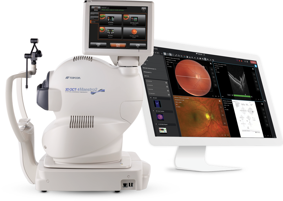
Topcon Maestro2 OCT instrument with review monitor

Our equipment at a glance

Test
Instrument we house
What it measures
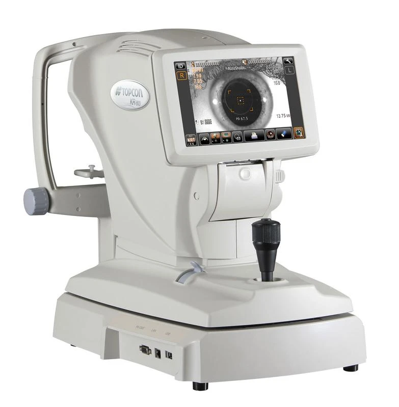
Autorefraction and keratometry
Topcon auto kerato-refractometer
Focusing power of the eye and corneal curvature
OCT and OCT angiography
ZEISS Cirrus OCT, Topcon Maestro2
Retinal layers, optic nerve and retinal blood vessels
Fundus photography
Topcon automatic fundus camera
True-color photographs of the retina and optic nerve
Visual field testing
ZEISS Humphrey Field Analyzer
Central and side vision
Optical biometry
Haag-Streit Lenstar
Length and internal dimensions of the eye
Corneal topography
Topcon Corneal Analyzer
Shape and curvature of the cornea
Corneal biomechanics and eye pressure
Reichert Ocular Response Analyzer
Eye pressure and how the cornea responds to it

Autorefraction and keratometry

  • What it is. Autorefraction is an automated measurement of how your eye focuses light. It gives an objective reading of nearsightedness, farsightedness and astigmatism. Keratometry, taken at the same time, measures the curvature of the cornea.
  • Why it matters. These readings are the starting point for understanding your vision. Because the instrument measures the same way every time, the numbers can be compared from visit to visit to show how your focusing power is changing.
  • What to expect. You look at a picture inside the instrument that drifts in and out of focus. Nothing touches your eye, and each eye takes only seconds.
Topcon auto kerato-refractometer

Topcon

Auto kerato-refractometer

A five-in-one instrument that pairs objective refraction and keratometry with checks of distance and near visual acuity, contrast sensitivity and vision under glare, all at one station.

Image & Information courtesy of Topcon.

Optical coherence tomography (OCT) and OCT angiography

  • What it is. OCT is a non-invasive scan that uses light to build cross-section images of the retina and optic nerve, layer by layer. OCT angiography goes a step further and maps the blood vessels in the retina without any dye injection.
  • Why it matters. These scans show the retina and optic nerve in fine detail, including thickness, swelling and the pattern of blood vessels. Repeating them over time lets us document how these structures change during your care.
  • What to expect. You rest your chin on a support and look at a target light. Nothing touches your eye, and the scan takes only moments per eye.
ZEISS Cirrus OCT instrument

ZEISS

Cirrus OCT

Our high-definition OCT for detailed scans of the macula, the retinal nerve fiber layer and the optic nerve head. It compares your results against earlier visits, so change over time is easy to see.

Image & Information courtesy of ZEISS.

Topcon Maestro2 OCT instrument with review monitor

Topcon

Maestro2 with OCT angiography

An automated instrument that captures an OCT scan and a true-color photograph of the back of the eye in one sitting. Its OCT angiography mode shows retinal blood flow without an injection.

Image & Information courtesy of Topcon.
Fundus photography

  • What it is. Fundus photography takes a true-color photograph of the back of your eye, showing the retina, the optic nerve head and the retinal blood vessels.
  • Why it matters. A photograph is a lasting visual record of how the back of your eye looks on a given day. Placing photographs from different visits side by side makes change over time easy to see.
  • What to expect. You look at a small target light and see a brief flash as the picture is taken. Nothing touches your eye, and the photograph can often be taken without dilating drops.
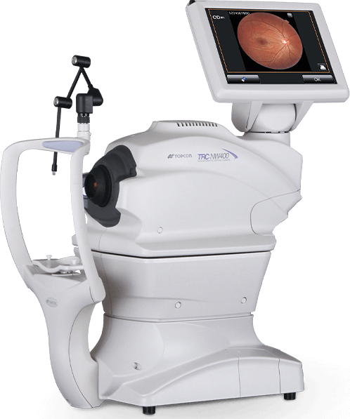
Topcon automatic fundus camera

Topcon

Automatic fundus camera

A non-mydriatic retinal camera that aligns, focuses and captures automatically. It photographs through small pupils with a low-intensity flash, and it can also photograph the front surface of the eye.

Image & Information courtesy of Topcon.

Visual field testing

  • What it is. A visual field test maps everything you can see while looking straight ahead, including your side vision.
  • Why it matters. Changes in side vision are easy to miss in daily life. This test records your field of vision objectively, so results can be compared from one visit to the next.
  • What to expect. You look into a bowl-shaped screen and press a button each time you see a small flash of light. One eye is tested at a time, and each eye takes a few minutes.
ZEISS Humphrey Field Analyzer

ZEISS

Humphrey Field Analyzer

The instrument most eye specialists rely on for automated perimetry. Its analysis software compares each test with your previous ones to show how your field of vision is changing.

Image & Information courtesy of ZEISS.

Optical biometry

  • What it is. Biometry measures the dimensions of your eye with light: its length, the curve of the cornea, and the depth and thickness of the structures in between.
  • Why it matters. The length of the eye and the size of its internal structures are key reference measurements. Recording them precisely gives us a baseline and lets us follow changes, such as lengthening of the eye, over time.
  • What to expect. You sit at the instrument and focus on a light for a few seconds. There is no contact with the eye and no need for numbing drops.
Haag-Streit Lenstar biometer with review monitor

Haag-Streit

Lenstar biometer

An optical biometer that records axial length, corneal curvature, corneal thickness, anterior chamber depth and lens thickness in a single measurement, giving a complete profile of the eye in one sitting.

Image & Information courtesy of Haag-Streit.
Corneal topography

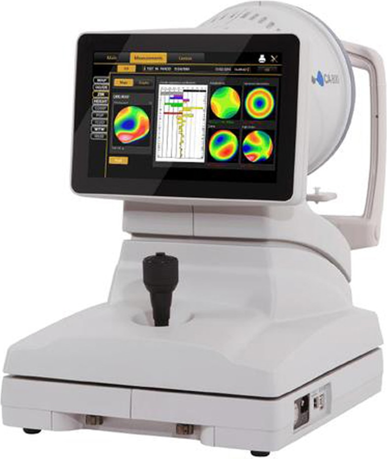
  • What it is. Topography creates a color-coded map of the shape of your cornea, the clear front window of the eye.
  • Why it matters. The shape of the cornea strongly affects how clearly you see. The map documents astigmatism and surface irregularities in detail and lets us compare your cornea from one visit to the next.
  • What to expect. You look at a pattern of lighted rings while the instrument takes a picture. It is quick, and nothing touches your eye.
Topcon Corneal Analyzer showing corneal maps on its screen

Topcon

Corneal Analyzer

A corneal topographer that maps the curvature of the front surface of the eye and displays the results as detailed color maps we can review with you on screen.

Image & Information courtesy of Topcon.
Corneal biomechanics and eye pressure

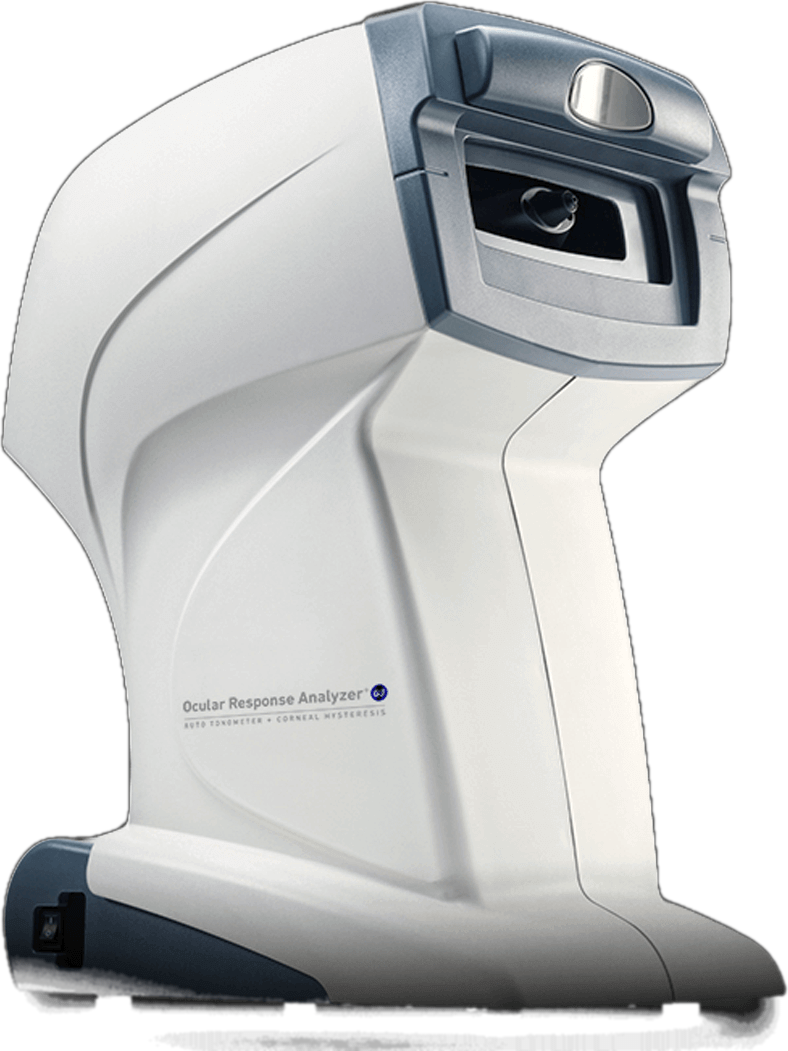
  • What it is. This test measures the pressure inside your eye and how your cornea responds to a gentle puff of air. That response is called corneal hysteresis.
  • Why it matters. The thickness and flexibility of the cornea affect ordinary pressure readings. Accounting for them gives a more reliable pressure value to record and follow over time.
  • What to expect. You feel a brief, soft puff of air on each eye. There is no contact and no drops are needed.
Reichert Ocular Response Analyzer

Reichert

Ocular Response Analyzer

A non-contact tonometer that measures corneal hysteresis and reports a cornea-compensated eye pressure, adding information that a standard pressure check does not provide.

Image & Information courtesy of Reichert.

Schedule a testing visit

We will let you know which of these tests are part of your evaluation. If you have questions about a test before your visit, our staff is glad to help.

Product names are trademarks of their respective manufacturers.