

Retinitis pigmentosa is a group of inherited retinal degenerations in which rod and then cone photoreceptors die progressively. Nothing in Ayurveda has been shown in a controlled trial to halt, slow, or reverse that loss, and no clinic anywhere can honestly promise restored sight. What genuinely helps is a molecular diagnosis, treatment of the complications that take extra vision on top of the disease, gene therapy when the genotype allows it, and skilled low-vision rehabilitation. Supportive care has a place in living with this condition. It is a modest place, and this page describes it plainly rather than dressing it up.
Complementary-care boundary: Ayurvedic eye care does not replace genetic testing and counseling, ophthalmic monitoring with optical coherence tomography and visual fields, carbonic anhydrase inhibitors or other treatment for cystoid macular edema, cataract surgery when the lens is limiting you, voretigene neparvovec gene therapy for biallelic RPE65 disease, or formal low-vision rehabilitation. We do not offer it as an alternative to any of these. If a practitioner suggests stopping your ophthalmology follow-up because a traditional protocol is working, that advice is wrong and it costs sight.
An Integrative Approach to the Drivers of Retinitis Pigmentosa
Families arrive at our New Jersey clinic having usually been told two things: that the diagnosis is genetic, and that there is no treatment. The first is accurate. The second is close enough to accurate that it should not be softened, but it is not the whole story. Retinitis pigmentosa is not one disease. More than eighty genes can cause it, inheritance may be autosomal dominant, autosomal recessive, X-linked, or mitochondrial, and the rate of loss differs enormously between those groups. A person with mild dominant disease may drive into their sixties. A child with an early recessive form may be registered blind before finishing school.
That variability matters because it changes what is worth doing. Knowing the causative gene tells you whether an approved therapy exists, whether a clinical trial is open, what to expect of the next decade, and what the risk is to siblings and children. Whole-exome and panel sequencing now return a causative variant in a substantial majority of affected people, with yield highest in consanguineous and well-characterized cohorts, and testing is the single most useful thing most patients have not yet done.
Alongside the genetics sits a second, more practical layer. A great deal of the vision people lose in retinitis pigmentosa is lost to things that are not the degeneration itself: posterior subcapsular cataract, cystoid macular edema, uncorrected refractive error, poor lighting, and untreated glare. These are addressable. Our integrative work concentrates there, on function, on systemic health, and on the exhaustion of living with a progressive condition, while the ophthalmic side of the plan does the parts that only ophthalmology can do.
Why Retinitis Pigmentosa Care Should Be Multi-Factorial
The Genotype Sets the Trajectory
Prognosis in retinitis pigmentosa tracks the gene far more closely than it tracks anything a patient does. X-linked RPGR disease tends to progress fast; some autosomal dominant rhodopsin variants progress slowly enough that useful reading vision persists for life. Without a molecular diagnosis, every conversation about the future is guesswork, and every supplement decision is made blind.
Two Separate Problems Run in Parallel
One is photoreceptor death, which current medicine cannot stop. The other is the collection of treatable complications that ride on top of it. Conflating the two leads people either to despair or to chase cures. Separating them lets us say honestly: this part we cannot change, and this part we can, and the second part is often worth several lines of acuity.
Cataract Arrives Early and Is Frequently Fixable
Posterior subcapsular cataract is far more common in retinitis pigmentosa than in the general population, and it arrives decades early. It scatters light, worsens glare, and blurs the small central island of vision people depend on. Removing it does not treat the retina, but it can meaningfully improve function in a patient who had assumed all further loss was irreversible.
Macular Edema Is Common and Treatable
Cystoid macular edema affects a substantial minority of eyes with retinitis pigmentosa and directly attacks the central vision that remains. It responds, at least partly, to oral carbonic anhydrase inhibitors, topical agents, and in selected eyes intravitreal therapy. A network meta-analysis of the available modalities found meaningful differences between them, which is precisely why this needs a retina specialist and not a supplement.
Syndromic Disease Hides Behind the Retina
Retinitis pigmentosa is sometimes the visible part of a systemic condition. Usher syndrome pairs it with sensorineural hearing loss. Bardet-Biedl involves obesity, kidney disease, and polydactyly. Refsum disease is driven by phytanic acid accumulation and responds to dietary restriction, which makes it one of the rare genuinely modifiable forms. Missing a syndrome means missing treatable systemic disease.
The Non-Visual Burden Is Part of the Disease
Losing the peripheral field costs independent mobility, night driving, employment, and often confidence long before central acuity falls. Rates of anxiety and depression in progressive retinal disease are high, and family dynamics shift when a genetic diagnosis implicates siblings and children. Care that addresses only the retina addresses perhaps half of what the condition actually does to a person.
Key Biological Mechanisms in Retinitis Pigmentosa
Rod Loss First, Cone Loss Second
Most causative genes are expressed in rods, so rods die first. That produces night blindness and midperipheral field loss. Cones then fail secondarily, even in patients whose mutation does not directly affect them, probably because rods supply trophic and metabolic support that cones cannot do without. Understanding this sequence explains why night vision goes years before reading vision.
Oxidative Stress in a High-Oxygen Retina
As rods die, oxygen consumption in the outer retina falls while choroidal supply continues, leaving surviving cones in a relatively hyperoxic environment. The resulting oxidative injury is one proposed driver of secondary cone death, and it is the rationale behind most antioxidant strategies. That rationale is biologically reasonable. It has not yet translated into a supplement with proven benefit on visual outcomes.
Retinal Pigment Epithelium and the Visual Cycle
The retinal pigment epithelium recycles retinoids so photoreceptors can regenerate light-sensitive pigment. When RPE65 is defective on both alleles, that cycle stalls and photoreceptors are starved of functional chromophore while remaining structurally present for a period. This narrow window is exactly what makes RPE65 disease the one form with an approved gene therapy.
Microglial Activation and Low-Grade Inflammation
Degenerating photoreceptors recruit microglia, which clear debris but also release inflammatory mediators that may accelerate the loss of neighboring cells. This is not inflammation in the sense of uveitis and it does not respond to the same drugs, but it is one reason anti-inflammatory approaches keep being studied. Evidence for clinical benefit remains preliminary.
Blood-Retinal Barrier Breakdown
Disruption of the inner and outer blood-retinal barriers allows fluid to accumulate in the macula. This is the mechanism behind cystoid macular edema in retinitis pigmentosa and is the reason carbonic anhydrase inhibitors, which enhance fluid transport across the retinal pigment epithelium, can reduce it. It is a mechanism with a real, testable treatment attached.
Inner Retinal Remodeling
Once photoreceptors are gone, the bipolar and ganglion cell layers do not simply wait. They rewire, sprout, and form aberrant connections. This remodeling is a major obstacle to any future restorative strategy, whether gene therapy, cell transplantation, or an electronic implant, and it is the strongest argument for intervening early rather than waiting for a promised cure.

How Retinitis Pigmentosa Presents and How It Progresses
Night Blindness Comes First
The earliest symptom is usually difficulty seeing in dim light and slow adaptation when moving from a bright room into a dark one. Children may be described as clumsy after sunset. Adults often date the change to a specific evening years earlier. Because the symptom is gradual and easily attributed to age, the average delay between first symptom and diagnosis is measured in years.
The Field Narrows From the Midperiphery Inward
Loss typically begins as a partial ring of reduced sensitivity in the midperipheral field, which enlarges and closes into a complete ring scotoma. The far periphery may persist for a time, giving a disconcerting pattern of a small central island plus a distant crescent. People bump into door frames and low obstacles while still reading fine print.
Central Acuity Often Holds for Decades
Many patients keep 20/25 to 20/40 central acuity well into middle age even when the field is severely constricted. This is why standard acuity testing alone badly underestimates disability, and why visual field and functional assessment matter more for decisions about driving, work, and mobility training.
Glare and Photophobia Are Underestimated
Bright sunlight, oncoming headlights, and fluorescent-lit supermarkets can be more disabling than dim conditions. Cone dysfunction, early cataract, and light scatter all contribute. Many people find good filters and hats more immediately useful than anything else offered to them.
Rate of Progression Varies by Inheritance
Longitudinal series show measurable annual decline in field and electroretinogram amplitude, with the steepest slopes in X-linked disease and the shallowest in some dominant forms. Averages are of limited use to an individual. What is useful is serial measurement in the same person on the same equipment over several years.
What Legal Blindness Means Here
In the United States, a visual field of twenty degrees or less in the better eye meets the definition of legal blindness regardless of acuity. Many people with retinitis pigmentosa qualify while still reading comfortably. Crossing that threshold opens access to rehabilitation services, assistive technology funding, and disability protections, and reaching it is not the same as losing all sight.
How Retinitis Pigmentosa Is Assessed at Netra Eye Institute
Assessment here is not a second opinion on your diagnosis. It is an attempt to establish exactly which form you have, how fast it is moving, which treatable complications are present right now, and which parts of your remaining function can be improved this year. Most of the following happens in coordination with your retina specialist and genetic counselor rather than in place of them.
Pedigree and Family History
We map three generations where possible, asking specifically about night vision, hearing, kidney disease, extra digits, and consanguinity. A well-taken pedigree frequently narrows the inheritance pattern before any test is ordered and identifies relatives who should be examined.
Dilated Examination and Wide-Field Imaging
Bone-spicule pigmentation, attenuated arterioles, and waxy disc pallor are the classic triad, but early disease can look almost normal. Wide-field color imaging documents the extent of pigmentary change and gives a reproducible baseline for comparison years later.
Optical Coherence Tomography
OCT measures the width of the surviving ellipsoid zone, which correlates with remaining cone function and is currently the most sensitive structural marker of progression. It also detects cystoid macular edema and epiretinal membrane, both of which are actionable findings.
Fundus Autofluorescence
A hyperautofluorescent ring around the fovea marks the boundary between functioning and failing retina. Tracking that ring as it constricts is one of the cleanest ways to follow the disease over time and is used as an endpoint in trials.
Full-Field Electroretinography
The ERG confirms generalized photoreceptor dysfunction, distinguishes rod-cone from cone-rod patterns, and detects disease before the fundus looks abnormal. In advanced cases responses are undetectable, at which point other measures carry the monitoring.
Visual Fields and Dark Adaptometry
Kinetic perimetry maps the remaining field better than standard automated testing in constricted eyes. Dark-adapted chromatic perimetry and dark adaptometry quantify the rod dysfunction patients actually experience, and both are used as outcome measures in gene therapy trials.
Genetic Testing and Counseling
Panel or whole-exome sequencing identifies a causative variant in most tested patients, and yield is highest when the phenotype is well characterized first. A result changes eligibility for approved therapy and trials, clarifies prognosis, and informs family planning. Uptake remains lower than it should be, and helping patients actually complete testing is part of the work.
Systemic and Nutritional Review
We check hearing, renal function where syndromic disease is suspected, and baseline liver function and lipid-soluble vitamin status before any supplement discussion. Women of childbearing potential need a documented pregnancy plan before vitamin A is ever considered.
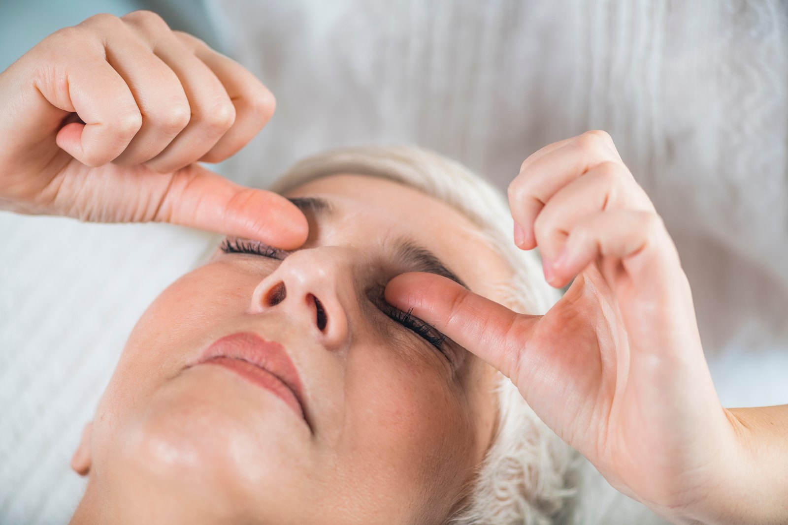
What Ayurvedic Eye Care Offers in Retinitis Pigmentosa
We should start with what the literature actually contains. There are no randomized controlled trials of Ayurvedic formulations, Netra Tarpana, Panchakarma, or any classical protocol showing an effect on visual field, electroretinogram amplitude, ellipsoid zone width, or acuity in retinitis pigmentosa. Surveys of patients with this diagnosis show that a large proportion try non-traditional and complementary treatments, and that reported benefit is overwhelmingly subjective. A small pilot study of an acupuncture protocol reported changes in some measures of visual function, but it was uncontrolled, tiny, and has not been replicated at a scale that would justify recommending it as treatment.
Given that, we offer supportive care under a narrow and stated remit. It is directed at symptoms, comfort, and the general health that surrounds a chronic eye condition, not at the degeneration. Every patient hears this sentence before anything is booked, and it appears in writing in the plan they take home.
- Ocular surface dryness and irritation, which are common and make glare and reading fatigue worse
- Eye strain from prolonged effort with a constricted field
- Sleep disruption, which is frequent when dim-light function is impaired and routines shift
- Stress, anxiety, and the low mood that often accompanies a progressive genetic diagnosis
- Dietary structure and adherence, built around whole foods rather than supplement marketing
- Digestive and metabolic health, which matters when systemic syndromes are involved
- Practical coaching around lighting, contrast, and pacing through the day
What it cannot do should be equally clear. It does not regenerate photoreceptors, restore lost visual field, improve electroretinogram responses, correct a genetic variant, or alter the rate of degeneration. Any practitioner who tells you otherwise is either mistaken or selling something, and in retinitis pigmentosa the cost of believing them is usually money, delay, and sometimes direct harm.
Where Ayurvedic Therapy Fits Alongside Genetic Diagnosis and Retinal Monitoring
After the Genetic Workup, Not Instead of It
We do not begin supportive care in a patient who has not had, or at least been offered, genetic testing. The molecular result determines whether an approved therapy or an open trial applies, and no supportive plan should be allowed to occupy the space where that conversation belongs.
Ophthalmic Follow-Up Continues Unchanged
Annual or biannual review with imaging and fields continues at whatever interval your retina specialist sets. If anything we do appeared to correlate with a change on those tests, the tests would still be the arbiter, not our impression.
Supplements Are Reviewed, Not Multiplied
Most patients arrive taking several products bought online. We list them, check doses against known thresholds, look for duplicated vitamin A, and usually remove more than we add. Stacking antioxidants is not a strategy.
Comfort Measures Use Clinic-Prepared Materials Only
Where an external therapy is offered, it uses sterile, clinic-prepared preparations with documented sourcing, applied to the periocular skin and lids. Nothing unregulated goes near the ocular surface, and no procedure is performed on an eye with active edema or recent surgery.
Treatable Complications Are Escalated Immediately
New central blur, distortion, or a step change in reading ability prompts same-week imaging, because cystoid macular edema and cataract both respond to treatment and both are missed when everything is attributed to the underlying disease.
Rehabilitation Is Arranged Early, Not Late
Orientation and mobility training, contrast and lighting assessment, magnification, and assistive technology are most useful before they become urgent. We refer at diagnosis rather than waiting for a crisis, and we treat that referral as a core part of treatment rather than a consolation.
What We Will Not Do
We will not use kajal, surma, or any unregulated traditional eye preparation, several of which have been shown to contain lead and other heavy metals. We will not recommend home-made eye washes, decoctions instilled into the eye, or unlabeled imported formulations. We will not administer or endorse intraocular stem-cell injections outside a registered trial, we will not claim to restore vision, and we will not advise anyone to delay or decline ophthalmic care.
Treatment Options, Honestly Compared
Vitamin A Palmitate
High-dose vitamin A palmitate is the most discussed intervention in retinitis pigmentosa, based on trial data suggesting a modest slowing of electroretinogram decline. A Cochrane review of vitamin A and fish oils concluded that the evidence does not establish a clear benefit on the outcomes patients care about, and later analyses of neuroprotective strategies reached similarly cautious conclusions. It is not a harmless supplement. It requires liver function and lipid monitoring, is contraindicated in pregnancy because of teratogenicity, and should be avoided in ABCA4-related disease, where retinoid loading is expected to worsen lipofuscin accumulation. Genotype first, then the discussion.
Omega-3 Fatty Acids and DHA
Ancillary analysis of the DHAX trial reported slower visual field progression in X-linked retinitis pigmentosa with docosahexaenoic acid supplementation, and observational work found higher omega-3 intake associated with better acuity outcomes in patients already taking vitamin A. These are secondary and observational findings, not proof. Dietary omega-3 intake is reasonable on general health grounds; high-dose supplementation is a decision to make with your retina specialist.
Lutein and Other Carotenoids
A randomized trial of lutein in patients receiving vitamin A found a small effect on midperipheral field with no effect on acuity or electroretinogram. Current reviews of nutritional supplements in retinitis pigmentosa and Stargardt disease describe the overall evidence as weak and inconsistent. Lutein is safe at dietary doses. That is the strongest honest statement available.
Voretigene Neparvovec for Biallelic RPE65 Disease
This is the one approved gene therapy, delivered by subretinal injection, and it applies only to patients with confirmed biallelic RPE65 mutations and sufficient viable retinal cells. Systematic reviews and post-approval data show real gains in dim-light navigation and light sensitivity, with acuity improvement more variable, alongside recognized risks including retinal thinning, chorioretinal atrophy, and the surgical hazards of subretinal delivery. Netra does not perform this procedure; we help identify candidates and refer.
Treating Cystoid Macular Edema
Oral carbonic anhydrase inhibitors such as acetazolamide, topical dorzolamide, and in some eyes intravitreal corticosteroid or anti-VEGF agents can reduce macular fluid. A network meta-analysis comparing modalities found differences in efficacy that matter when choosing, and recurrence after stopping is common. This is retina specialist territory and one of the few places where treating aggressively protects real function.
Cataract Surgery, Prostheses, and Unproven Cell Therapy
Cataract extraction can improve function in selected eyes, though posterior capsule opacification and zonular weakness are more common here. The Argus II retinal prosthesis produced measurable but crude visual percepts over five years in profound vision loss and is no longer commercially supported. Against that stands the clearest warning on this page: intravitreal injection of autologous adipose-derived cells at unregulated clinics has caused severe, permanent vision loss, including in patients who had useful sight beforehand. Cell therapy for inherited retinal disease remains investigational, and the only safe route into it is a registered trial.
Building a Daily Plan Around Retinitis Pigmentosa
Start From the Genetic Report
The plan changes depending on the gene. ABCA4 disease means avoiding vitamin A loading. Refsum disease means phytanic acid restriction, which genuinely alters the course. RPE65 disease means an urgent referral conversation. Building habits before knowing the genotype risks building the wrong ones.
Do Not Self-Prescribe Vitamin A
Doses used in trials are far above dietary intake and carry hepatic and teratogenic risk. If vitamin A is used at all it belongs under supervision with baseline and periodic liver function testing, a clear pregnancy plan, and a check that no other product in the cabinet also contains retinol.
Eat for the Retina Without Chasing a Bottle
Oily fish two or three times a week, dark leafy greens, colored vegetables, nuts, and olive oil deliver carotenoids and omega-3 fatty acids in the matrix they occur in. This is sound general advice with modest specific evidence, and it is cheaper and safer than the supplement shelf.
Manage Light Rather Than Avoiding It
Amber or plum tinted filters reduce glare and can improve contrast for many patients; a wide-brimmed hat does more than most people expect. Ultraviolet protection is prudent, though it has not been shown to slow the degeneration. Indoors, task lighting placed behind the shoulder beats a brighter ceiling.
Protect the Vision You Still Have
Polycarbonate protective lenses for sports and workshop tasks matter more when the reserve is small. Stopping smoking is worth doing on retinal and systemic grounds. Blood pressure, glucose, and lipids still need attention, because a second retinal disease on top of this one is a genuinely bad outcome.
Set Up the Home for a Narrow Field
Contrast markings on stair edges, consistent furniture placement, decluttered walkways, and even lighting without deep shadows prevent the falls that a constricted field makes likely. Orientation and mobility training teaches scanning techniques that no supplement can substitute for.
Look After the Rest of Your Life
Sleep, exercise, and social contact hold up function and mood. Talk to an employer early about accommodation rather than late about failure. Peer organizations and genetic counseling help families work out what the diagnosis means for siblings and children, and that conversation is often the one people postpone longest.
Red Flags That Need Urgent Care
Retinitis pigmentosa progresses slowly. Anything sudden is not the degeneration and should be treated as a separate, possibly treatable, event. Contact an ophthalmologist or emergency service the same day if any of the following occurs.
- A sudden shower of new floaters, flashing lights, or a curtain across part of the field, which may indicate retinal detachment
- Rapid loss of central vision over days rather than years
- New distortion of straight lines, suggesting macular edema or membrane
- Eye pain, redness, or a fixed mid-dilated pupil with halos, which can indicate acute angle closure
- Any vision change following intraocular surgery or injection
- Vision loss in one eye only, since retinitis pigmentosa is characteristically symmetrical
- New double vision, facial weakness, or severe headache accompanying visual change
- Rapidly worsening night vision in an adult with no family history, which can reflect autoimmune or paraneoplastic retinopathy
- Vision change after starting a new medication, particularly hydroxychloroquine or thioridazine
Several conditions mimic retinitis pigmentosa, including drug toxicity, prior infection, and cancer-associated retinopathy. If your diagnosis was made without an electroretinogram and genetic testing, it is reasonable to ask for both before accepting it as final.
Frequently Asked Questions on Retinitis Pigmentosa
No. There is no controlled trial evidence that any Ayurvedic treatment halts, slows, or reverses retinitis pigmentosa, and no mechanism by which oils, herbal preparations, or Panchakarma would replace dead photoreceptors. Clinics advertising cures for this condition are marketing to families in distress. Supportive care can help with comfort, dryness, sleep, and coping. It cannot restore sight.
Not outside a registered clinical trial. Intravitreal injection of autologous adipose-derived cells at unregulated clinics has caused severe and permanent vision loss, including in eyes that had useful vision before the procedure. Legitimate cell therapy research for inherited retinal disease exists, is early, and never asks patients to pay large sums for an unapproved injection. If a clinic charges you for it, that is the clearest warning sign.
Only after genetic testing and only with a physician monitoring you. The benefit is uncertain, the Cochrane assessment was cautious, and the risks are real: liver injury with long-term high doses, and serious harm to a fetus. In ABCA4-related disease it is specifically avoided. This is not a supplement to start on your own.
Yes, for four practical reasons. It determines eligibility for the approved RPE65 gene therapy and for open trials. It refines prognosis. It identifies syndromic forms with treatable systemic features. And it tells your siblings and children what their own risk is. Modern sequencing finds a causative variant in most people tested.
When performed with sterile, clinic-prepared material on an eye with no active edema, recent surgery, or surface infection, it is generally well tolerated as a comfort measure. It is not a treatment for the retina and we do not present it as one. Unregulated preparations and traditional eye cosmetics such as kajal and surma are a different matter entirely and have been shown to contain lead.
Selected References for Scientific Support
- Hartong DT, Berson EL, Dryja TP. Retinitis pigmentosa. Lancet. 2006. PubMed
- Schwartz SG, Wang X, Chavis P, et al. Vitamin A and fish oils for preventing the progression of retinitis pigmentosa. Cochrane Database Syst Rev. 2020. PubMed
- Hoffman DR, Hughbanks-Wheaton DK, Spencer R, et al. Docosahexaenoic acid slows visual field progression in X-linked retinitis pigmentosa: ancillary outcomes of the DHAX trial. Invest Ophthalmol Vis Sci. 2015. PubMed
- Berson EL, Rosner B, Sandberg MA, et al. Omega-3 intake and visual acuity in patients with retinitis pigmentosa receiving vitamin A. Arch Ophthalmol. 2012. PubMed
- Berson EL, Rosner B, Sandberg MA, et al. Clinical trial of lutein in patients with retinitis pigmentosa receiving vitamin A. Arch Ophthalmol. 2010. PubMed
- Barthelemy N, Lee W, Gregori NZ, et al. Nutritional supplements: current evidence for retinitis pigmentosa and Stargardt disease. Curr Opin Ophthalmol. 2026. PubMed
- Sherratt-Mayhew S, Bisp M, Waheed S, et al. A systematic review of neuroprotective therapies for visual acuity preservation in retinitis pigmentosa. BMJ Open Ophthalmol. 2026. PubMed
- Butt AB, Zaman M, Mihalache A, et al. Voretigene neparvovec (Luxturna) for biallelic RPE65-associated retinal dystrophy: systematic review. Am J Ophthalmol. 2026. PubMed
- Li JXL, Rowson AC, Naji L, et al. Post-approval outcomes of voretigene neparvovec retinal gene therapy: a systematic review and meta-analysis. Surv Ophthalmol. 2026. PubMed
- Nagiel A, Russell SR, Lam BL, et al. Interim results from a multicenter observational safety study of patients treated with voretigene neparvovec-rzyl in the United States. Ophthalmology. 2026. PubMed
- Jaju S, Sivagnanam J, Menon P, et al. Whole-exome sequencing in a consanguinity-enriched South Indian retinitis pigmentosa cohort: diagnostic yield and molecular spectrum. Ophthalmol Sci. 2026. PubMed
- Kuriyan AE, Albini TA, Townsend JH, et al. Vision loss after intravitreal injection of autologous stem cells for AMD. N Engl J Med. 2017. PubMed
- Huang Y, Xie Y, Wang C. Stem cell-based therapies for inherited retinal diseases: translational advances and clinical evidence. Biomol Biomed. 2026. PubMed
- Yang HA, Jheng WL, Hwang YS, et al. Comparative efficacy of different treatment modalities in the management of macular edema in retinitis pigmentosa: a systematic review and network meta-analysis. BMJ Open Ophthalmol. 2026. PubMed
- da Cruz L, Dorn JD, Humayun MS, et al. Five-year safety and performance results from the Argus II retinal prosthesis system clinical trial. Ophthalmology. 2016. PubMed
- Bittner AK, Gould JM, Rosenfarb A, et al. A pilot study of an acupuncture protocol to improve visual function in retinitis pigmentosa patients. Clin Exp Optom. 2014. PubMed
- Kiser AK, Dagnelie G. Reported effects of non-traditional treatments and complementary and alternative medicine by retinitis pigmentosa patients. Clin Exp Optom. 2008. PubMed
- Saper RB, Phillips RS, Sehgal A, et al. Lead, mercury, and arsenic in US- and Indian-manufactured Ayurvedic medicines sold via the Internet. JAMA. 2008. PubMed
- Mukhopadhyay S, Abraham SE, Holla B, et al. Heavy metals in Indian traditional systems of medicine: a systematic scoping review and recommendations for integrative medicine practice. J Altern Complement Med. 2021. PubMed