

Keratoconus thins and steepens the cornea, and the single most important thing you can change about its course is whether you rub your eyes. Eye rubbing is the dominant modifiable driver of progression, which means any therapy that involves pressure, massage, or manipulation around the eye is not neutral here; it works in the same direction as the disease. Corneal cross-linking is the only treatment shown to halt progression. Ayurveda has no trial evidence in keratoconus at all. What supportive care can honestly do is attack the itch and the habit that put hands on the cornea in the first place.
Complementary-care boundary: Ayurvedic eye care does not replace corneal collagen cross-linking, rigid gas permeable or scleral contact lenses, intrastromal corneal ring segments, topography-guided surface ablation, deep anterior lamellar keratoplasty, or penetrating keratoplasty. It does not replace serial corneal tomography, the test that determines whether your cornea is progressing. Nothing offered at Netra Eye Institute substitutes for any of these. A cornea that is progressing and not cross-linked keeps getting worse, and the scarring and irregularity that follow are not reversible.
An Integrative Approach to the Drivers of Keratoconus
Keratoconus is a bilateral, usually asymmetric ectasia. The corneal stroma loses structural collagen organization, thins focally, and bulges forward into a cone. Vision degrades not because the eye needs a stronger prescription but because the optical surface becomes irregular, and irregularity cannot be corrected by a spherocylindrical lens. That is why patients describe ghosting, streaking, and glare long before the letters on the chart get smaller.
The disease has a genetic substrate that nobody can change and a mechanical driver that many people can. Repeated, forceful eye rubbing deforms an already weakened cornea, raises corneal temperature, and releases inflammatory enzymes into the tear film. Systematic review and meta-analysis have consistently linked rubbing to keratoconus, and the association strengthens with the duration and force of rubbing. Behind most persistent rubbing sits an itch, usually allergic. Treat the itch properly and the rubbing often stops.
This is where integrative care has something real to contribute, and it is narrower than it sounds. Netra Eye Institute works alongside your cornea specialist, not in place of one. We do not perform cross-linking, we do not fit the lenses that carry your vision, and we do not decide when you need surgery. What we do is help with allergy, sleep, ocular surface comfort, and the stubborn behavioral work of leaving your eyes alone.
Why Keratoconus Care Should Be Multi-Factorial
Eye rubbing is the driver you can actually change
Genetics load the gun, and rubbing is the part under your control. Meta-analysis of case-control data finds a consistent association between habitual rubbing and keratoconus, and the risk scales with how long and how hard people rub. Cases of strikingly asymmetric disease often track with a dominant rubbing hand or a preferred sleeping side. This is not a minor lifestyle footnote. It is the central behavioral target of the whole condition.
Allergy and atopy sit behind the rubbing
Allergic eye disease is associated with keratoconus in systematic review, and the mechanism is not mysterious: itchy eyes get rubbed. Vernal keratoconjunctivitis in children is a particularly high-risk combination, since the itch is severe and the cornea is young and elastic. Controlling allergy is therefore corneal treatment, not just symptom relief.
Progression is fastest when patients are youngest
Keratoconus usually declares itself in the teens or twenties and progresses most aggressively in that window. A pediatric diagnosis is a different clinical situation from a diagnosis at forty, and specialists often move to cross-linking sooner in younger patients rather than waiting to document change. Time matters here in a way it does not in most eye conditions.
Biomechanics, not just shape
Topographic steepening is a late signal of a mechanical problem that started earlier. The cornea loses stiffness before it visibly changes shape, which is why biomechanical measurement and posterior elevation maps can flag disease that front-surface curvature still calls normal. Thinking in terms of structural weakness rather than curvature explains why a physical insult like rubbing matters so much.
Acuity does not capture the disability
A patient can read 20/25 on a high-contrast chart and still be unable to drive at night, read a whiteboard, or tolerate oncoming headlights. Higher-order aberrations, monocular diplopia, and glare are the real burden, and studies of vision-related quality of life in keratoconus show substantial impact that a letter score does not reflect.
The uncertainty is its own problem
Being told you have a progressive corneal disease in your twenties, then asked to come back in six months to find out whether it moved, is genuinely difficult. Patients delay lens fittings, avoid appointments, or chase alternative cures out of fear. Naming that pressure and answering questions honestly is part of care, and it is one reason we refuse to sell false hope on this page.
Key Biological Mechanisms in Keratoconus
Disorganized collagen and focal thinning
The healthy cornea holds its shape through hundreds of collagen lamellae woven in a specific architecture, with interweaving concentrated anteriorly. In keratoconus that organization degrades, lamellae slip past one another, and the stroma thins in a localized zone. Once enough structural collagen is lost, normal intraocular pressure is sufficient to push the weakened area outward.
What rubbing does mechanically
Rubbing applies direct shear and compression to a cornea that has lost stiffness. Measurements during vigorous rubbing show transient spikes in intraocular pressure and a rise in ocular surface temperature. Repeated thousands of times over years, this is a chronic mechanical loading pattern applied precisely where the tissue is weakest. The link is biologically plausible and epidemiologically consistent.
Proteases and inflammatory mediators
Tears from keratoconic eyes show elevated matrix metalloproteinase-9 and inflammatory cytokines, and levels rise further with rubbing and allergic disease. These enzymes degrade extracellular matrix. Keratoconus was long described as non-inflammatory, and that description has not survived the tear film data. It behaves more like a condition with a low-grade inflammatory component layered onto a structural weakness.
Keratocyte loss
Keratocytes maintain and repair stromal matrix. Their density is reduced in keratoconic corneas, particularly in the thinned zone, which impairs the tissue's ability to rebuild what enzymes and mechanical stress remove. Less repair capacity plus more degradation is a poor combination in a tissue that must stay optically precise.
Genetic susceptibility
Family history raises risk several-fold, and keratoconus is more common in Down syndrome and in some connective tissue disorders. Genome-wide studies have identified multiple loci influencing corneal thickness and curvature. Genetics explains why two people who rub their eyes equally do not end up with the same cornea, and it is the part of the picture no intervention modifies.
Descemet membrane rupture and hydrops
If the cornea thins and stretches far enough, Descemet membrane can tear. Aqueous floods into the stroma, the cornea swells dramatically, and vision drops over hours to days with pain and light sensitivity. This is acute corneal hydrops. It usually settles over weeks but frequently leaves a dense scar, and it is more common in eyes that keep being rubbed.

How Keratoconus Presents and How It Progresses
The early years look like a bad prescription
A teenager or young adult needs new glasses unusually often, and each new pair helps less than the last. Astigmatism climbs and its axis shifts. Night driving becomes unpleasant first. Many patients pass through several optometrists before someone orders corneal tomography, which is the test that actually makes the diagnosis.
Established disease and its signs
As the cone develops, examination shows a scissoring retinoscopic reflex, stromal thinning, fine vertical stress lines in the deep stroma known as Vogt striae, and an iron deposition ring at the cone base. Corneal nerves become unusually visible. In advanced disease the lower lid takes on a V shape when the patient looks down.
Acute hydrops
Sudden clouding of the cornea with pain, tearing, and marked light sensitivity signals a break in Descemet membrane. It is not an infection, but it needs same-week specialist care. Treatment is supportive, occasionally with intracameral gas, and the swelling resolves over weeks. The residual scar often determines whether a transplant becomes necessary.
Both eyes, unequally
Keratoconus is essentially always bilateral when looked for carefully with tomography, even when one eye seems normal. The asymmetry is often striking, and it frequently corresponds to sleeping side or rubbing hand. A truly unilateral case should prompt a careful search for asymmetric mechanical trauma.
Most disease stabilizes eventually
Progression typically slows through the thirties and forties as corneal collagen naturally cross-links with age. That is genuine reassurance for an older patient with stable scans. It is not reassurance for a nineteen-year-old, who may have fifteen years of potential change ahead and needs active monitoring.
Ectasia after refractive surgery
A similar process can follow LASIK or PRK when the cornea was too thin, too irregular, or subclinically ectatic before surgery. It presents the same way, progresses the same way, and is treated with cross-linking and specialty lenses in the same way. Screening tomography before refractive surgery exists to prevent exactly this.
How Keratoconus Is Assessed at Netra Eye Institute
Your cornea specialist owns the diagnosis, the tomography series, and the decision about cross-linking. We read those results with you, look hard at the factors that drive progression, and make sure nothing we suggest works against your treatment. If you have been diagnosed and are not under specialist follow-up, arranging that is the first thing we do.
A detailed history of rubbing
We ask specifically and without judgment: which hand, how hard, how often, with knuckles or fingertips, on waking or through the day, and whether it follows an itch. Many patients have never been asked and do not realize how much they do it. This history is often the most useful information in the whole visit.
Refraction and best-corrected acuity
We record spectacle and, where relevant, lens-corrected acuity, along with how the prescription has moved over recent years. A rapidly shifting cylinder axis carries more information than a single number. We also ask what you cannot do comfortably, since that is what treatment is meant to fix.
Corneal tomography and its trend
Tomography maps the front surface, the back surface, and thickness across the whole cornea. Posterior elevation change and the thinnest pachymetry point often shift before front-surface curvature does. We review your serial scans as a series, because measurement variability easily imitates progression on any two scans compared in isolation.
What counts as progression
Consensus guidance defines progression by consistent change in more than one parameter, typically steepening of the anterior surface, change in posterior elevation, and thinning, rather than by a single index crossing a threshold. Knowing the criteria helps patients understand why one steeper reading is not automatically a reason for treatment, and why two are.
Corneal biomechanics
Where available, dynamic corneal response measurement adds information about stiffness that shape alone does not provide. It is most useful in borderline cases and in screening the apparently normal fellow eye. We treat it as supporting evidence rather than a standalone diagnosis.
Allergy and ocular surface examination
We look at the tarsal conjunctiva for papillae, assess tear film stability and meibomian gland function, and ask about seasonal patterns, pets, dust, and personal or family atopy. Elevated tear inflammatory markers and allergic surface disease both sit on the pathway between itch and corneal damage.
Contact lens review
If you wear rigid or scleral lenses, we check comfort, wearing hours, solution use, and whether a poor fit is causing the surface irritation that makes you rub. Lens problems belong with your fitter, and we refer back rather than adjusting anything ourselves.
Systemic and family context
We ask about Down syndrome, connective tissue disorders, sleep apnea, and the sleeping position you actually use. First-degree relatives of keratoconus patients should be screened with tomography, especially siblings and children, because early detection is what makes timely cross-linking possible.
What Ayurvedic Eye Care Offers in Keratoconus
Start with the plain finding: a search of the published literature returns no clinical trials of Ayurvedic treatment in keratoconus. Not a small trial, not a weak one, none. There is no evidence that any oil therapy, herbal formulation, or purification protocol flattens a cone, thickens a cornea, improves corneal biomechanics, or slows progression. We are not going to describe that void as promising.
What supportive care can do in this condition is indirect and genuinely useful. The pathway from allergy to itch to rubbing to corneal damage is the best-supported modifiable chain in keratoconus, and most of that chain responds to ordinary, careful management. Helping a patient stop rubbing is a corneal intervention delivered through behavior rather than through the eye, and it costs nothing and risks nothing.
- Structured work on stopping eye rubbing, including trigger identification, substitution strategies, and follow-up that treats the habit as a clinical target
- Allergy management in coordination with your physician, so that the itch driving the rubbing is actually treated
- Ocular surface and dry eye care, since a dry, gritty surface produces the urge to rub
- Sleep review, including sleeping position and pressure on one eye through the night
- Support for tolerating rigid and scleral lenses, and prompt referral back to your fitter when the problem is the lens
- A safety review of every eye preparation, supplement, and home remedy you currently use
- Help reading your tomography reports so that monitoring feels comprehensible rather than alarming
What it cannot do needs stating without hedging. Ayurvedic therapy does not reverse corneal thinning, does not flatten the cone, does not restore corneal collagen, does not remove scarring, and does not substitute for cross-linking, specialty contact lenses, or transplantation. Choosing it instead of cross-linking in a progressing cornea means allowing damage that no later treatment undoes.
Where Ayurvedic Therapy Fits Alongside Cross-Linking and Specialty Lenses
The cross-linking decision comes first
Before anything supportive is discussed, we want to know whether your cornea is progressing and what your specialist has advised. If cross-linking has been recommended and you are hesitating, our job is to help you understand it and go ahead, not to offer an alternative. A cornea that progresses while you deliberate does not wait for you.
Breaking the rubbing habit
Treating rubbing as a habit rather than a moral failing is what makes it stoppable. We map when it happens, which is usually on waking, at screens, and during allergy season, and build substitutions: cold compress instead of knuckles, a deliberate blink, artificial tears within reach, fingernails kept short. Patients who quit rubbing often notice their scans settle, and that is the outcome worth chasing.
Treating the itch so the hands stay down
Willpower alone fails against a severe allergic itch. Allergen reduction at home, cool compresses, and physician-directed antihistamine or mast cell stabilizing drops address the cause. In children with vernal disease this is urgent corneal care, not cosmetic relief, and it belongs with an allergist or ophthalmologist rather than with herbal preparations.
Why we will not massage, press, or manipulate around the eye
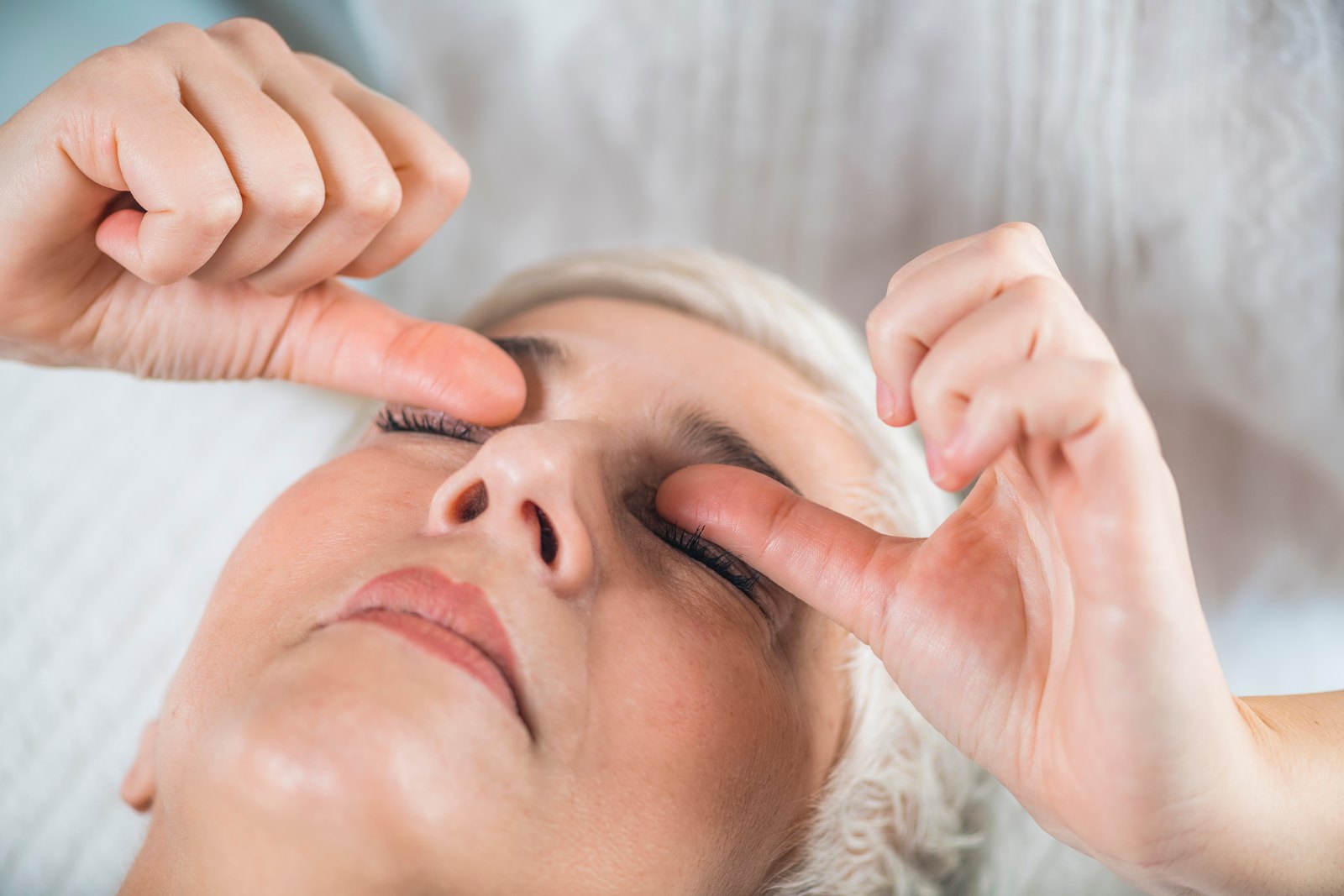
This is the clearest safety boundary on this page. A keratoconic cornea has lost stiffness, and external pressure deforms it. Since repeated mechanical loading is the leading modifiable driver of progression, any therapy built on pressure is working with the disease rather than against it. That rules out periocular massage, marma or acupressure points on and around the globe, firm lid manipulation, eye exercises that involve pressing, and any technique a practitioner describes as improving circulation by applying pressure to the eye. We do not offer them in keratoconus, and we ask patients receiving them elsewhere to stop.
Netra Tarpana: restricted, and never in an unstable cornea
Netra Tarpana pools warm ghee within a dough ring around the orbit. It has no evidence in keratoconus and is never presented as treatment for it. Where it is considered at all, it is for surface dryness in a cornea documented as stable, with sterile technique, no pressure on the globe, and never in an eye with recent hydrops, active inflammation, a corneal graft, or a healing epithelium after cross-linking.
Coordination with your cornea specialist
We write to your specialist describing what we are doing and why, and we ask for your tomography reports. If your specialist wants something stopped, it stops. Supportive care that runs in parallel and unreported is not integrative care; it is a second plan nobody is supervising.
What We Will Not Do
We will not use kajal, surma, or any unregulated traditional eye preparation. These have repeatedly been found to contain lead, and testing of Ayurvedic medicines sold in the United States and India has detected lead, mercury, or arsenic in a substantial share of samples. We will not recommend home-made eye washes, decoctions, or rinses, which add infection and chemical injury risk to an already fragile surface. We will not apply pressure or massage to a keratoconic eye, will not advise delaying or declining cross-linking, will not discourage rigid or scleral lens wear, and will not claim that anything we provide reverses corneal thinning or removes the need for surgery.
Treatment Options, Honestly Compared
Glasses and soft lenses
In mild disease, spectacles or soft toric lenses may give usable vision. They correct regular astigmatism only, so as irregularity grows they stop working, and increasing the prescription does not help. Specialized soft lenses designed for keratoconus extend this stage somewhat. Neither affects progression.
Rigid gas permeable and scleral lenses
A rigid surface replaces the irregular cornea optically, and for most patients with moderate disease this is what restores functional vision. Scleral lenses vault the cornea entirely and rest on the sclera, which suits sensitive or very irregular eyes and adds a fluid reservoir that helps dryness. Fitting is specialized and iterative. These lenses correct vision; they do not stop the cornea from changing.
Corneal collagen cross-linking
Riboflavin plus ultraviolet-A light creates additional bonds between stromal collagen fibrils, stiffening the tissue. It is the only treatment shown to halt progression, and United States multicenter clinical trial data supported its approval for progressive keratoconus and post-refractive ectasia. It is stabilizing, not restorative: most eyes hold steady, some flatten modestly, and vision is not expected to improve substantially. Recovery involves several days of discomfort after epithelial removal, and infection and haze are uncommon but real risks. Netra Eye Institute does not perform cross-linking and refers for it.
Intrastromal ring segments
Small arcs placed within the corneal stroma flatten and regularize the cone, often improving lens tolerance and reducing aberrations. More recently, segments made from donor corneal tissue have been used similarly. They are a shape-modifying option for eyes that cannot tolerate lenses, usually combined with cross-linking, and results vary with cone type and position.
Topography-guided surface ablation with cross-linking
In selected stable or simultaneously cross-linked eyes, a shallow, topography-guided laser treatment can smooth the irregular surface. Tissue removal in an already thin cornea is not a decision to take lightly, candidacy is narrow, and the goal is better lens tolerance and reduced aberration rather than freedom from correction.
Corneal transplantation
When scarring or extreme irregularity defeats every lens, a graft is the remaining option. Deep anterior lamellar keratoplasty replaces the diseased stroma while retaining the patient's own endothelium, which lowers rejection risk; penetrating keratoplasty replaces the full thickness. Meta-analysis comparing the two in keratoconus shows broadly similar visual outcomes with a better endothelial safety profile for the lamellar approach. Recovery takes a year or more, and most patients still need a contact lens afterward.
Building a Daily Plan Around Keratoconus
Do not rub, and have something else ready
The instruction to stop rubbing only works if something replaces it. Keep preservative-free artificial tears where your hands go: bedside, desk, car, bag. When the urge arrives, instill a drop and blink firmly several times. If you must touch, press a cool compress flat against a closed lid without moving it. Never grind with knuckles.
Morning is the highest-risk moment
Most rubbing happens in the first minute after waking, often before you are properly conscious of it. A drop of lubricant within reach of the bed, splashing the face on rising, and a short cool compress interrupt the reflex. Patients who fix this one moment often cut their total rubbing sharply.
Take allergy seriously all year
Wash bedding hot, keep windows closed in pollen season, rinse hair before bed, and keep pets out of the bedroom. Use the antihistamine or mast cell stabilizing drops your physician prescribes consistently through the season rather than only on bad days. Cool compresses relieve itch without mechanical loading.
Sleep position and pressure on the eye
Sleeping face down or hard on one side presses the orbit into the pillow for hours. Asymmetric keratoconus often matches the favored side. Sleeping on your back is the safest option, and a firmer pillow or a positional wedge helps if you tend to roll. If you snore heavily or wake unrefreshed, ask about sleep apnea testing.
Lens wear and hygiene
Follow your fitter's schedule for wearing hours and replacement, use only the solutions recommended, and never rinse lenses with tap water. Discomfort is information rather than something to tolerate, since an uncomfortable lens leads straight back to rubbing. Report redness, pain, or blur with lens wear the same day.
Screens, dryness, and blinking
Blink rate falls sharply at a screen, and a dry surface itches. Take a genuine break every twenty minutes or so, blink completely rather than halfway, reposition the monitor below eye level to reduce exposed surface area, and keep the air from vents off your face.
Keep the scan appointments
Progression is defined by change across serial tomography, so missed scans mean progression is detected late. If you are young, recently diagnosed, or newly cross-linked, the interval your specialist chose is deliberate. Book the next appointment before leaving the clinic.
Red Flags That Need Urgent Care
Most keratoconus changes slowly enough to be managed at scheduled visits. The exceptions below do not wait, and none of them should be treated with a supportive therapy while you see whether things settle. When in doubt, contact your cornea specialist or an emergency eye service the same day.
- Sudden clouding of vision in one eye with pain, tearing, and strong light sensitivity, which suggests acute corneal hydrops.
- Any pain, redness, discharge, or blurring while wearing rigid or scleral lenses, which can mean corneal infection and needs assessment the same day.
- A white or gray spot visible on the cornea, particularly with pain or light sensitivity.
- Rapid change in prescription or acuity over weeks rather than months, in a patient who has not been re-scanned.
- Redness, pain, or falling vision in an eye that has had a corneal graft, since these can signal rejection and early treatment matters.
- Worsening pain, haze, or blur in the weeks after cross-linking, rather than the expected steady improvement.
- Severe, unrelenting itch that you cannot stop yourself rubbing, which is an emergency for the cornea even though it feels minor.
- A first-degree relative newly diagnosed while you have never had tomography, especially if you are under thirty.
- Any injury or blow to an eye with keratoconus or a corneal graft.
- New double vision in one eye that persists when the other is covered, if it is a change from your usual pattern.
Keratoconus itself does not blind people overnight. Infection, hydrops, and graft rejection can cause damage quickly, and all three are treatable when seen early. Being seen unnecessarily costs an afternoon; waiting can cost a cornea.
Frequently Asked Questions on Keratoconus
No. There are no clinical trials of Ayurvedic treatment in keratoconus, and nothing in the literature shows that any formulation or therapy thickens a thinned cornea, flattens a cone, or restores corneal collagen. Corneal cross-linking is the only treatment shown to halt progression. Claims of reversal describe hope, not evidence.
Yes. It is the strongest modifiable driver we know of. Systematic review and meta-analysis link habitual rubbing to keratoconus, the association strengthens with how long and how forcefully people rub, and asymmetric disease often matches a dominant hand or a preferred sleeping side. Stopping costs nothing and is the single most valuable thing you can do yourself.
No, and we will not provide them. A keratoconic cornea has lost stiffness, so external pressure deforms it, and repeated mechanical loading is exactly what drives progression. Periocular massage, acupressure on the globe, firm lid manipulation, and pressing eye exercises all push in the wrong direction. If someone is offering these for your cornea, stop and tell your specialist.
No. If your cornea is documented as progressing, cross-linking is what stops it, and delaying means accepting thinning and irregularity that no later treatment undoes. Stopping rubbing and controlling allergy are worth doing alongside it, not instead of it. That decision belongs to your cornea specialist.
No, and we refuse to use them. Kajal and surma have repeatedly been found to contain lead, and testing of Ayurvedic medicines sold in the United States and India has detected lead, mercury, or arsenic in a large share of samples. Home-made washes add contamination and chemical injury risk on a cornea that is already thin. Bring what you are using and we will review it.
Selected References for Scientific Support
- Gomes JA, Tan D, Rapuano CJ, et al. Global consensus on keratoconus and ectatic diseases. Cornea. 2015. PubMed
- Gomes JAP, Hafezi F, Ambrosio R Jr, et al. Global Consensus on Keratoconus and Ectatic Diseases-Edition 2. Cornea. 2026. PubMed
- Hashemi H, Heydarian S, Hooshmand E, et al. The Prevalence and Risk Factors for Keratoconus: A Systematic Review and Meta-Analysis. Cornea. 2020. PubMed
- Sahebjada S, Al-Mahrouqi HH, Moshegov S, et al. Eye rubbing in the aetiology of keratoconus: a systematic review and meta-analysis. Graefes Arch Clin Exp Ophthalmol. 2021. PubMed
- Seth I, Bulloch G, Vine M, et al. The association between keratoconus and allergic eye diseases: A systematic review and meta-analysis. Clin Exp Ophthalmol. 2023. PubMed
- Song M, Fang QY, Seth I, et al. Non-genetic risk factors for keratoconus. Clin Exp Optom. 2023. PubMed
- Ren S, Tu R, Xu L, et al. A high body mass index strengthens the association between the time of eye rubbing and keratoconus in a Chinese population: a case control study. BMC Public Health. 2023. PubMed
- Belin MW, Alizadeh R, Torres-Netto EA, et al. Determining Progression in Ectatic Corneal Disease. Asia Pac J Ophthalmol (Phila). 2020. PubMed
- Hersh PS, Stulting RD, Muller D, et al. United States Multicenter Clinical Trial of Corneal Collagen Crosslinking for Keratoconus Treatment. Ophthalmology. 2017. PubMed
- Greenstein SA, Hersh PS. Corneal Crosslinking for Progressive Keratoconus and Corneal Ectasia: Summary of US Multicenter and Subgroup Clinical Trials. Transl Vis Sci Technol. 2021. PubMed
- Gustafsson I, Olafsdottir T, Neumann O, et al. A randomised controlled trial on cross-linking protocols for the treatment of progressive keratoconus using isoosmolar and hypoosmolar riboflavin with the addition of sterile water in thin corneae. Acta Ophthalmol. 2026. PubMed
- Awad AA, Elettreby AM, Abo Elnaga AA, et al. Penetrating Keratoplasty versus Deep Anterior Lamellar Keratoplasty for Keratoconus: A Systematic Review and Meta-Analysis of 27,018 Eyes. Semin Ophthalmol. 2025. PubMed
- Kanu LN, Boychev N, Fry M, et al. Sociodemographic predictors of acute corneal hydrops in patients with unstable keratoconus. PLoS One. 2025. PubMed
- Ben-Shaul O, Schwartz S, Segal A, et al. Factors Associated with Pediatric Keratoconus - A Population-Based Nested Case-Control Study. Ophthalmic Epidemiol. 2026. PubMed
- Alsalamah S, Abosabaah Y, Alhabs G, et al. Behavioral and Sociodemographic Factors Associated with Vision-Related Quality of Life in Keratoconus: A Cross-Sectional Study in Riyadh, Saudi Arabia. Vision (Basel). 2026. PubMed
- Mazzotta C, Gagliano C, D'Esposito F, et al. Anti-inflammatory and dry eye benefits of accelerated epi-off corneal cross-linking in pediatric keratoconus with allergic ocular surface disease and elevated MMP-9. Eye Vis (Lond). 2026. PubMed
- Saper RB, Phillips RS, Sehgal A, et al. Lead, mercury, and arsenic in US- and Indian-manufactured Ayurvedic medicines sold via the Internet. JAMA. 2008. PubMed
- Mukhopadhyay S, Abraham SE, Holla B, et al. Heavy Metals in Indian Traditional Systems of Medicine: A Systematic Scoping Review and Recommendations for Integrative Medicine Practice. J Altern Complement Med. 2021. PubMed