
Search conditions, treatments, research reviews and articles. Press Enter to see all results, Esc to close.
Search conditions, treatments, research reviews and articles. Press Enter to see all results, Esc to close.

Blog
July 30, 2026
Corneal neuralgia is pain coming from damaged corneal nerves rather than from a dry or inflamed surface — which is why it persists when the examination looks normal and drops do not help. It can follow LASIK, infection, injury or chronic dry eye. This page covers how it is recognized and how it is managed.
Laser-assisted in situ keratomileusis (LASIK) has transformed refractive surgery, offering millions of patients freedom from spectacles and contact lenses. While the procedure is highly successful and safe, a small but significant subset of patients develops a debilitating condition known as post-LASIK corneal neuropathic pain (CNP)—a chronic pain disorder arising from damage to the corneal nerves.
Unlike typical postoperative dry eye or surface irritation, neuropathic corneal pain is characterized by persistent, severe ocular discomfort that is often disproportionate to clinical findings, making diagnosis and treatment particularly challenging.
In recent years, growing evidence suggests that traditional treatments—primarily focused on lubrication or anti-inflammatory strategies—are insufficient for many of these patients. This has led to increasing interest in regenerative and integrative therapies, such as Netra Restoration Therapy, which aim to address the underlying neurobiological dysfunction rather than merely suppress symptoms.
LASIK involves creation of a corneal flap, which inevitably transects corneal nerves. This leads to:
Patients with neuropathic pain show reduced corneal nerve fiber density and increased neuromas, as seen in in vivo confocal microscopy studies.
Corneal neuropathic pain is defined as pain resulting from dysfunction or damage to the corneal somatosensory nervous system. It manifests with symptoms such as:
Dry eye, infection, trauma, recurrent erosion, contact-lens injury, toxic exposure, surgery including LASIK, and systemic small-fiber neuropathy can disturb corneal nerves. Repeated epithelial stress may sustain nociceptive input, while injured fibers may develop abnormal sprouts, altered ion channels, or ectopic firing. Dry eye, neurotrophic keratopathy, recurrent erosion, and neuropathic pain can overlap, but they are not interchangeable diagnoses.
Tear instability and epithelial stress activate keratocytes, immune cells, cytokines, proteases, and neuromediators. These signals can lower sensory thresholds. Peripheral sensitization may then produce hyperalgesia or allodynia to light, air, touch, or temperature. Persistent trigeminal input may promote central or nociplastic amplification, allowing symptoms to continue after the surface has improved. No single biomarker confirms corneal neuralgia.
Corneal neuralgia may follow ocular-surface disease, infection, trauma, epithelial injury, surgery, or systemic neuropathy. Migraine, fibromyalgia or other overlapping pain syndromes, autoimmune or metabolic disease, sleep disturbance, anxiety, depression, and chronic pain history may increase vulnerability or amplify disability. Ocular surgery is one of several possible initiating events, but it does not define the condition. These are associations, not proof of causation.
Examination by your ophthalmologist should assess tear breakup, staining, lid and meibomian disease, corneal sensitivity, and neurotrophic change. A topical anesthetic challenge may suggest a predominantly peripheral component when pain improves, or central contribution when pain persists; it is not definitive. In vivo confocal microscopy may show reduced or abnormal nerves, microneuromas, or increased dendritic cells; findings must be interpreted with the history and examination.
A corneal specialist should exclude infection, exposure, recurrent erosion, ectasia, herpetic disease, and neurotrophic keratopathy. Care may include preservative-free lubricants, meibomian treatment, anti-inflammatory therapy, autologous serum or platelet-rich plasma tears, scleral or PROSE lenses, and neuropathic-pain medication. Neurology, pain medicine, or behavioral health may help when central sensitization, migraine, sleep disturbance, or impaired function is prominent.
Standard management includes:
However, these approaches often fail because:
This creates a need for therapies that target neuro-regeneration and functional recovery.
Netra Restoration Therapy (NRT) is based on an integrative approach combining:
Unlike conventional treatments, NRT aims to restore the structural and functional integrity of the corneal neurovascular unit.
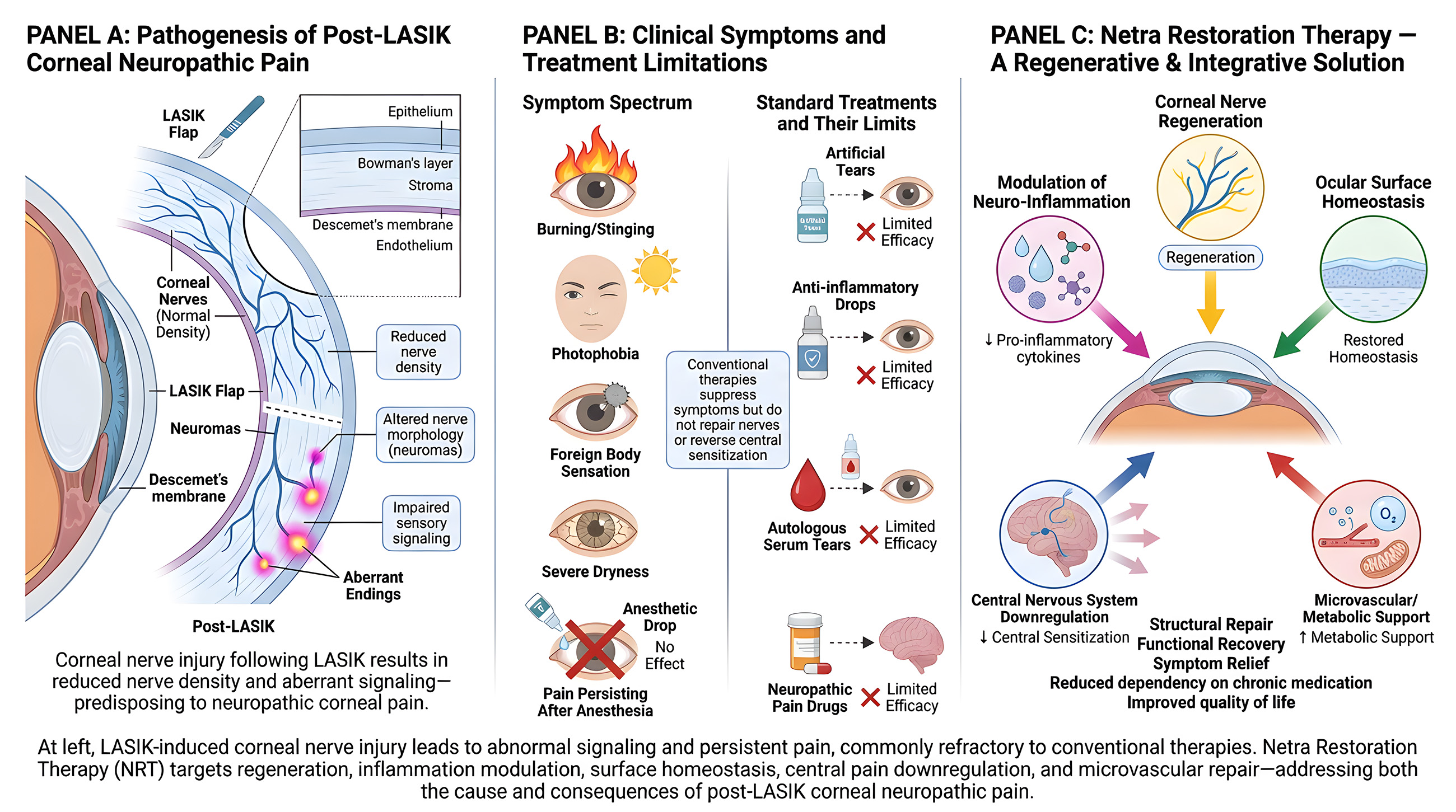
One of the central goals of NRT is promoting nerve regeneration:
Given that reduced nerve density is a key driver of neuropathic pain , regenerative approaches directly address the root cause.
NRT incorporates therapies that:
This helps interrupt the chronic inflammatory loop responsible for persistent pain.
By improving:
NRT reduces peripheral triggers that perpetuate nerve sensitization.
Through sustained peripheral healing and neuromodulatory approaches:
This is critical in chronic cases where pain persists independent of ocular surface pathology.
Emerging evidence suggests that microvascular dysfunction contributes to nerve pathology. NRT protocols often include:
These factors are essential for nerve repair and long-term stability.
Post-LASIK neuropathic pain requires multimodal treatment—a fact emphasized in clinical studies.
NRT inherently provides:
By addressing root causes, NRT may:
Patients with neuropathic corneal pain often suffer severe psychological and functional impairment. Effective restoration therapy can:
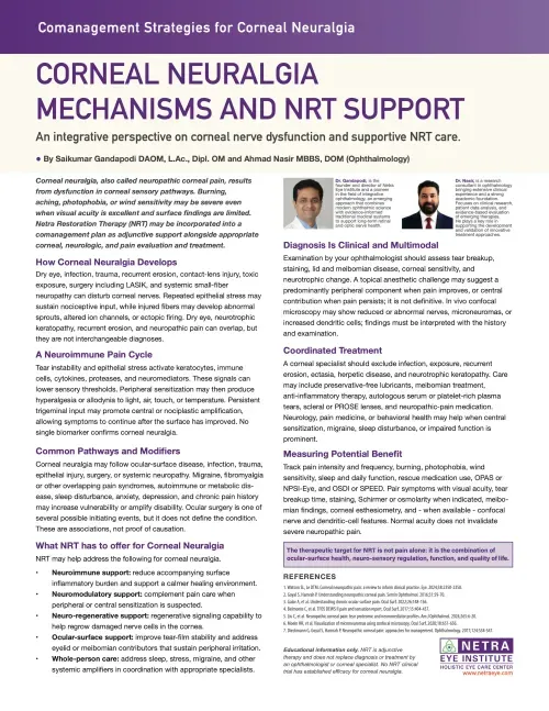
Download: Corneal Neuralgia — Mechanisms and NRT Support (PDF)
Educational information only. NRT is adjunctive therapy and does not replace diagnosis or treatment by an ophthalmologist or corneal specialist. No NRT clinical trial has established efficacy for corneal neuralgia.
Post-LASIK corneal neuropathic pain represents a complex and often misunderstood complication of refractive surgery. Rooted in corneal nerve injury, neuro-inflammation, and central sensitization, it cannot be effectively managed by traditional surface-based therapies alone.
Netra Restoration Therapy offers a promising solution by:
As our understanding of corneal neurobiology continues to expand, therapies like NRT that focus on true restoration rather than symptomatic relief are likely to become central to the management of this challenging condition.



